Lung volumes and emphysema in smokers with interstitial lung abnormalities
- PMID: 21388308
- PMCID: PMC3074462
- DOI: 10.1056/NEJMoa1007285
Lung volumes and emphysema in smokers with interstitial lung abnormalities
Abstract
Background: Cigarette smoking is associated with emphysema and radiographic interstitial lung abnormalities. The degree to which interstitial lung abnormalities are associated with reduced total lung capacity and the extent of emphysema is not known.
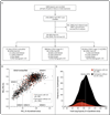
Methods: We looked for interstitial lung abnormalities in 2416 (96%) of 2508 high-resolution computed tomographic (HRCT) scans of the lung obtained from a cohort of smokers. We used linear and logistic regression to evaluate the associations between interstitial lung abnormalities and HRCT measurements of total lung capacity and emphysema.
Results: Interstitial lung abnormalities were present in 194 (8%) of the 2416 HRCT scans evaluated. In statistical models adjusting for relevant covariates, interstitial lung abnormalities were associated with reduced total lung capacity (-0.444 liters; 95% confidence interval [CI], -0.596 to -0.292; P<0.001) and a lower percentage of emphysema defined by lung-attenuation thresholds of -950 Hounsfield units (-3%; 95% CI, -4 to -2; P<0.001) and -910 Hounsfield units (-10%; 95% CI, -12 to -8; P<0.001). As compared with participants without interstitial lung abnormalities, those with abnormalities were more likely to have a restrictive lung deficit (total lung capacity <80% of the predicted value; odds ratio, 2.3; 95% CI, 1.4 to 3.7; P<0.001) and were less likely to meet the diagnostic criteria for chronic obstructive pulmonary disease (COPD) (odds ratio, 0.53; 95% CI, 0.37 to 0.76; P<0.001). The effect of interstitial lung abnormalities on total lung capacity and emphysema was dependent on COPD status (P<0.02 for the interactions). Interstitial lung abnormalities were positively associated with both greater exposure to tobacco smoke and current smoking.
Conclusions: In smokers, interstitial lung abnormalities--which were present on about 1 of every 12 HRCT scans--were associated with reduced total lung capacity and a lesser amount of emphysema. (Funded by the National Institutes of Health and the Parker B. Francis Foundation; ClinicalTrials.gov number, NCT00608764.).
Conflict of interest statement
No other potential conflict of interest relevant to this article was reported.
Figures


Comment in
-
Smoking and subclinical interstitial lung disease.N Engl J Med. 2011 Mar 10;364(10):968-70. doi: 10.1056/NEJMe1013966. N Engl J Med. 2011. PMID: 21388315 No abstract available.
-
Smokers with interstitial lung abnormalities.N Engl J Med. 2011 Jun 23;364(25):2465-6; author reply 2466. doi: 10.1056/NEJMc1104014. N Engl J Med. 2011. PMID: 21696313 No abstract available.
-
Smokers with interstitial lung abnormalities.N Engl J Med. 2011 Jun 23;364(25):2465; author reply 2466. doi: 10.1056/NEJMc1104014. N Engl J Med. 2011. PMID: 21696314 No abstract available.
-
MUC5B and pulmonary fibrosis, omalizumab for severe allergic asthma, and interstitial lung abnormalities in smokers with emphysema.Am J Respir Crit Care Med. 2012 May 1;185(9):1021-2. doi: 10.1164/rccm.201110-1846RR. Am J Respir Crit Care Med. 2012. PMID: 22550211 No abstract available.
References
-
- Smoking and health: a report of the Advisory Committee to the Surgeon General of the Public Health Service. Washington, DC: Public Health Service; 1964. (PHS publication no. 1103.)
-
- Webb WR. Thin-section CT of the secondary pulmonary lobule: anatomy and the image — the 2004 Fleischner lecture. Radiology. 2006;239:322–338. - PubMed
Publication types
MeSH terms
Associated data
Grants and funding
- UL1 TR000005/TR/NCATS NIH HHS/United States
- G0701127/MRC_/Medical Research Council/United Kingdom
- U01 HL089897/HL/NHLBI NIH HHS/United States
- 5R21CA116271-2/CA/NCI NIH HHS/United States
- K12 HD052892/HD/NICHD NIH HHS/United States
- K08 HL092222/HL/NHLBI NIH HHS/United States
- UL1 TR000454/TR/NCATS NIH HHS/United States
- K23 HL087030/HL/NHLBI NIH HHS/United States
- R21 CA116271/CA/NCI NIH HHS/United States
- K23 HL089353/HL/NHLBI NIH HHS/United States
- K25 HL104085/HL/NHLBI NIH HHS/United States
- T32 HL07427/HL/NHLBI NIH HHS/United States
- T32 HL007427/HL/NHLBI NIH HHS/United States
- R01 HL111024/HL/NHLBI NIH HHS/United States
- U01 HL089856/HL/NHLBI NIH HHS/United States
LinkOut - more resources
Full Text Sources
Other Literature Sources
Medical
Research Materials
