Case Reports
. 2011 Mar 10;364(10):939-46.
doi: 10.1056/NEJMoa1006939.

A dystroglycan mutation associated with limb-girdle muscular dystrophy

Affiliations
Case Reports

A dystroglycan mutation associated with limb-girdle muscular dystrophy

Yuji Hara et al. N Engl J Med. .

Abstract

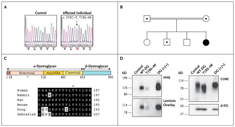
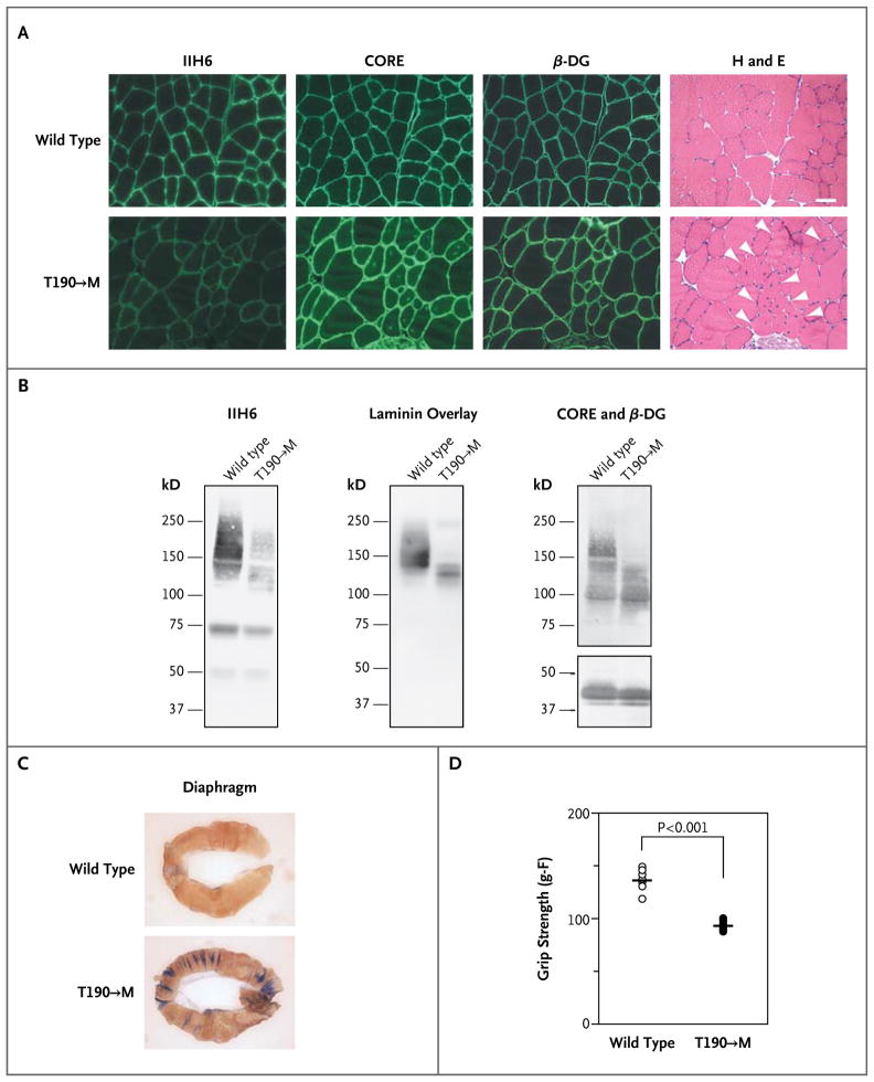
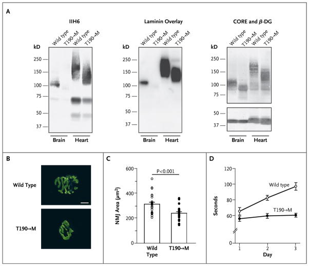
Dystroglycan, which serves as a major extracellular matrix receptor in muscle and the central nervous system, requires extensive O-glycosylation to function. We identified a dystroglycan missense mutation (Thr192→Met) in a woman with limb-girdle muscular dystrophy and cognitive impairment. A mouse model harboring this mutation recapitulates the immunohistochemical and neuromuscular abnormalities observed in the patient. In vitro and in vivo studies showed that the mutation impairs the receptor function of dystroglycan in skeletal muscle and brain by inhibiting the post-translational modification, mediated by the glycosyltransferase LARGE, of the phosphorylated O-mannosyl glycans on α-dystroglycan that is required for high-affinity binding to laminin.


Figures

Figure 1
Figure 1. Identification of a DAG1 Missense Mutation in a Woman with Limb-Girdle Muscular Dystrophy, and In Vitro Biochemical Characterization of the Encoded Protein
In Panel A, electrochromatograms reveal sequence variation in DAG1. A homozygous mutation, c.575C→T (arrow), leads to an amino acid change, T192→M. The T192→M variant was not detected in any of the 100 Turkish control subjects tested (200 chromosomes). Panel B shows the family pedigree. Squares denote male family members, and circles female family members; the double line indicates a consanguineous marriage; the black circle denotes the affected woman, and dots indicate heterozygous carriers. Panel C shows a schematic representation of a dystroglycan mutant protein (top). Human α-dystroglycan is composed of a signal peptide (SP) (amino acids 1 through 29), an N-terminal domain (amino acids 30 through 316), a mucinlike domain (amino acids 317 through 485), and a C-terminal domain (amino acids 486 through 653). The position of the patient’s mutation (T192→M) is indicated by an asterisk. Amino acid residues are aligned flanking the codon affected by the T192→M missense mutation (bottom). Among these residues, black boxes denote identity and gray boxes similarity. Panel D shows a biochemical analysis of wild-type (WT-DG) and T192→M dystroglycan in dystroglycan-null myoblast cells. Functional modification of α-dystroglycan was evaluated by Western blotting with an antibody (IIH6) that recognizes only the glycosylated form of α-dystroglycan and by laminin-overlay assay. Overall expression of wild-type dystroglycan and T192→M dystroglycan was detected with the use of antibodies against the α-dystroglycan protein core (CORE) and antibodies against β-dystroglycan (β-DG).
Figure 2
Figure 2. Biochemical and Physiological Skeletal-Muscle Phenotypes of Disease in Gene-Targeted Mice with the Same Dystroglycan Mutation as the Patient
Panel A shows immunofluorescence and histologic analyses of T190→M skeletal-muscle (iliopsoas) sections prepared when the animals were 21 weeks of age. Serial sections were stained with antibodies against glycosylated α-dystroglycan (IIH6), the α-dystroglycan core (CORE), and β-dystroglycan (β-DG). Histologic abnormalities in the sections were evaluated by means of hematoxylin and eosin (H and E) staining. White arrowheads indicate centrally nucleated fibers (scale bar, 50 μm). Panel B shows a representative biochemical analysis of wild-type (WT) and T190→M α-dystroglycan isolated from wheat-germ agglutinin–enriched homogenates of skeletal muscle. Both Western blotting and laminin-overlay assays were carried out. Functional modification of α-dystroglycan was impaired by the T190→M mutation in vivo, as shown by the dramatic reduction in IIH6 immunoreactivity and laminin binding in the absence of major differences in CORE and β-DG immunoreactivity. Panel C shows the uptake of Evans blue dye in the diaphragm after exercise in wild-type and T190→M mice at 8 weeks of age. Panel D shows the whole-mouse grip-strength measurements, in gram-force (g–F), for the T190→M mice and their wild-type littermates. The P value was obtained with the use of Student’s t-test.
Figure 3
Figure 3. Neuromuscular Phenotypes in Mice with the Dystroglycan Mutation
Panel A shows functional modification of α-dystroglycan in the brain and heart of mice with the T190→M mutation. Antibody staining and laminin-overlay assays were performed on blots of wheat-germ agglutinin–enriched homogenates from the brain and heart of T190→M mice and their wild-type (WT) littermates. Panels B and C show defects in maturation of the neuromuscular junction (NMJ) in the T190→M mice. Panel B shows representative images of the NMJ in the diaphragm; specimens were stained, with α-Bungarotoxin labeled with Alexa Fluor 488 (scale bar, 10 μm). Panel C shows individual data (circles) and mean (±SE) values for the NMJ area in wild-type and T190→M mice. Panel D shows the mean (±SE) values of five rotating-rod trials performed to assess neurologic defects in the T190→M mice. The time that the mice remained on the rotating rod was significantly longer for the wild-type mice than for the T190→M mice (P<0.001 by one-way analysis of variance).

References

    1. Ibraghimov-Beskrovnaya O, Ervasti JM, Leveille CJ, Slaughter CA, Sernett SW, Campbell KP. Primary structure of dystrophin-associated glycoproteins linking dystrophin to the extracellular matrix. Nature. 1992;355:696–702. - PubMed
    1. Barresi R, Campbell KP. Dystroglycan: from biosynthesis to pathogenesis of human disease. J Cell Sci. 2006;119:199–207. - PubMed
    1. Michele DE, Barresi R, Kanagawa M, et al. Post-translational disruption of dystroglycan-ligand interactions in congenital muscular dystrophies. Nature. 2002;418:417–22. - PubMed
    1. Beltrán-Valero de Bernabé D, Currier S, Steinbrecher A, et al. Mutations in the O-mannosyltransferase gene POMT1 give rise to the severe neuronal migration disorder Walker-Warburg syndrome. Am J Hum Genet. 2002;71:1033–43. - PMC - PubMed
    1. van Reeuwijk J, Janssen M, van den Elzen C, et al. POMT2 mutations cause α-dystroglycan hypoglycosylation and Walker-Warburg syndrome. J Med Genet. 2005;42:907–12. - PMC - PubMed

Publication types