

A novel copper complex induces paraptosis in colon cancer cells via the activation of ER stress signalling
- PMID: 21388518
- PMCID: PMC3823100
- DOI: 10.1111/j.1582-4934.2011.01292.x
A novel copper complex induces paraptosis in colon cancer cells via the activation of ER stress signalling
Abstract
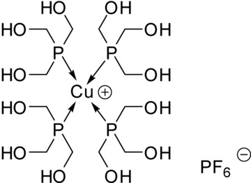
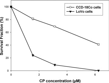
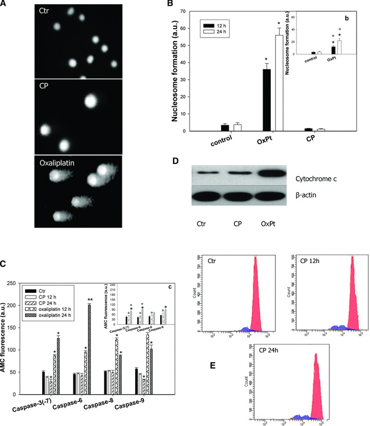
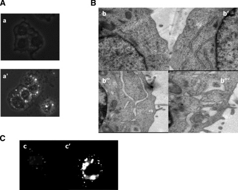
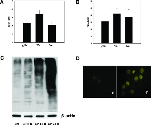
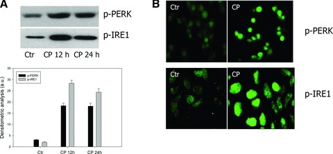
Platinum anticancer drugs have been used for three decades despite their serious side effects and the emerging of resistance phenomena. Recently, a phosphine copper(I) complex, [Cu(thp)(4)][PF(6)] (CP), gained special attention because of its strong antiproliferative effects. CP killed human colon cancer cells more efficiently than cisplatin and oxaliplatin and it overcame platinum drug resistance. CP preferentially reduced cancer cell viability whereas non-tumour cells were poorly affected. Colon cancer cells died via a programmed cell death whose transduction pathways were characterized by the absence of hallmarks of apoptosis. The inhibition of 26S proteasome activities induced by CP caused intracellular accumulation of polyubiquitinated proteins and the functional suppression of the ubiquitin-proteasome pathway thus triggering endoplasmic reticulum stress. These data, providing a mechanistic characterization of CP-induced cancer cell death, shed light on the signaling pathways involved in paraptosis thus offering a new tool to overcome apoptosis-resistance in colon cancer cells.
© 2011 The Authors Journal of Cellular and Molecular Medicine © 2011 Foundation for Cellular and Molecular Medicine/Blackwell Publishing Ltd.
Figures
Similar articles
-
Hollow Calcium/Copper Bimetallic Amplifier for Cuproptosis/Paraptosis/Apoptosis Cancer Therapy via Cascade Reinforcement of Endoplasmic Reticulum Stress and Mitochondrial Dysfunction.ACS Nano. 2024 Oct 29;18(43):30053-30068. doi: 10.1021/acsnano.4c11455. Epub 2024 Oct 16. ACS Nano. 2024. PMID: 39412236
-
Calreticulin is a fine tuning molecule in epibrassinolide-induced apoptosis through activating endoplasmic reticulum stress in colon cancer cells.Mol Carcinog. 2017 Jun;56(6):1603-1619. doi: 10.1002/mc.22616. Epub 2017 Feb 16. Mol Carcinog. 2017. PMID: 28112451
-
The thioxotriazole copper(II) complex A0 induces endoplasmic reticulum stress and paraptotic death in human cancer cells.J Biol Chem. 2009 Sep 4;284(36):24306-19. doi: 10.1074/jbc.M109.026583. Epub 2009 Jun 26. J Biol Chem. 2009. PMID: 19561079 Free PMC article.
-
Novel copper complexes as potential proteasome inhibitors for cancer treatment (Review).Mol Med Rep. 2017 Jan;15(1):3-11. doi: 10.3892/mmr.2016.6022. Epub 2016 Dec 9. Mol Med Rep. 2017. PMID: 27959411 Review.
-
Exploring paraptosis as a therapeutic approach in cancer treatment.J Biomed Sci. 2024 Nov 4;31(1):101. doi: 10.1186/s12929-024-01089-4. J Biomed Sci. 2024. PMID: 39497143 Free PMC article. Review.
Cited by
-
Organometallic Palladium Complexes with a Water-Soluble Iminophosphorane Ligand as Potential Anticancer Agents.Organometallics. 2012 Aug 27;31(16):5772-5781. doi: 10.1021/om3006239. Epub 2012 Jul 25. Organometallics. 2012. PMID: 23066172 Free PMC article.
-
Therapeutic potential of the phosphino Cu(I) complex (HydroCuP) in the treatment of solid tumors.Sci Rep. 2017 Oct 24;7(1):13936. doi: 10.1038/s41598-017-13698-1. Sci Rep. 2017. PMID: 29066771 Free PMC article.
-
Unveiling the Potential of Innovative Gold(I) and Silver(I) Selenourea Complexes as Anticancer Agents Targeting TrxR and Cellular Redox Homeostasis.Chemistry. 2022 Dec 15;28(70):e202201898. doi: 10.1002/chem.202201898. Epub 2022 Oct 25. Chemistry. 2022. PMID: 36106679 Free PMC article.
-
Knowledge mapping of copper-induced cell death: A bibliometric study from 2012 to 2022.Medicine (Baltimore). 2022 Nov 11;101(45):e31133. doi: 10.1097/MD.0000000000031133. Medicine (Baltimore). 2022. PMID: 36397452 Free PMC article.
-
Endoplasmic reticulum stress signalling - from basic mechanisms to clinical applications.FEBS J. 2019 Jan;286(2):241-278. doi: 10.1111/febs.14608. Epub 2018 Aug 4. FEBS J. 2019. PMID: 30027602 Free PMC article. Review.
References
-
- Kartalou M, Essigmann JM. Mechanisms of resistance to cisplatin. Mutat Res. 2001;478:23–43. - PubMed
-
- Zhang CX, Lippard SJ. New metal complexes as potential therapeutics. Curr Opin Chem Biol. 2003;7:481–9. - PubMed
-
- Simon TM, Kunishima DH, Vibert GJ, et al. Screening trial with the coordinated gold compound auranofin using mouse lymphocyte leukemia P388. Cancer Res. 1981;41:94–7. - PubMed
-
- Berners-Price SJ, Johnson RK, Mirabelli CK, et al. Copper(I) complexes with bidentate tertiary phosphine ligands: solution chemistry and antitumor activity. Inorg Chem. 1987;26:3383–7.
-
- Berners-Price SJ, Mirabelli CK, Johnson RK, et al. In vivo antitumor activity and in vitro cytotoxic properties of bis [1,2-bis(diphenylphosphino)ethane]gold(I) chloride. Cancer Res. 1986;46:5486–9. - PubMed
Publication types
MeSH terms
Substances
LinkOut - more resources
Full Text Sources
Other Literature Sources
Miscellaneous
