. 2011 Mar 9:10:25.
doi: 10.1186/1476-4598-10-25.

Examination of thromboxane synthase as a prognostic factor and therapeutic target in non-small cell lung cancer

Affiliations

Examination of thromboxane synthase as a prognostic factor and therapeutic target in non-small cell lung cancer

Mary-Clare Cathcart et al. Mol Cancer. .

Abstract

Background: Thromboxane synthase (TXS) metabolises prostaglandin H2 into thromboxanes, which are biologically active on cancer cells. TXS over-expression has been reported in a range of cancers, and associated with a poor prognosis. TXS inhibition induces cell death in-vitro, providing a rationale for therapeutic intervention. We aimed to determine the expression profile of TXS in NSCLC and if it is prognostic and/or a survival factor in the disease.

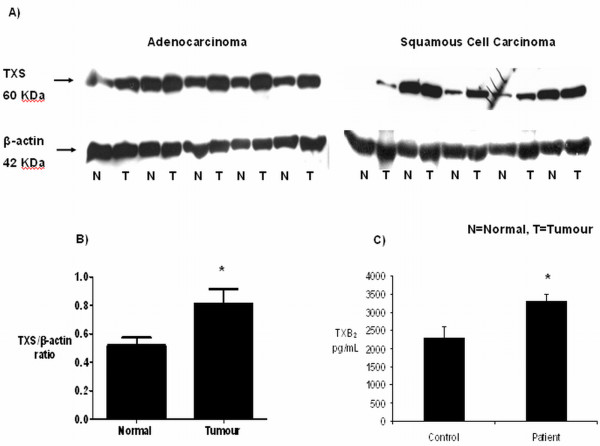
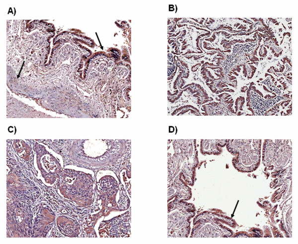
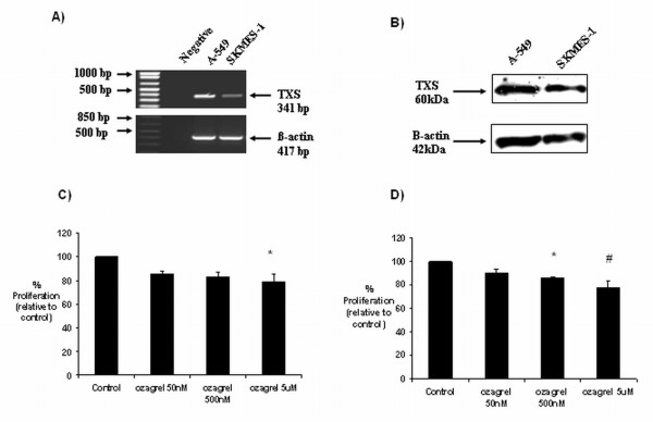
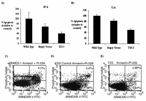
Methods: TXS expression was examined in human NSCLC and matched controls by western analysis and IHC. TXS metabolite (TXB2) levels were measured by EIA. A 204-patient NSCLC TMA was stained for COX-2 and downstream TXS expression. TXS tissue expression was correlated with clinical parameters, including overall survival. Cell proliferation/survival and invasion was examined in NSCLC cells following both selective TXS inhibition and stable TXS over-expression.

Results: TXS was over-expressed in human NSCLC samples, relative to matched normal controls. TXS and TXB2 levels were increased in protein (p < 0.05) and plasma (p < 0.01) NSCLC samples respectively. TXS tissue expression was higher in adenocarcinoma (p < 0.001) and female patients (p < 0.05). No significant correlation with patient survival was observed. Selective TXS inhibition significantly reduced tumour cell growth and increased apoptosis, while TXS over-expression stimulated cell proliferation and invasiveness, and was protective against apoptosis.

Conclusion: TXS is over-expressed in NSCLC, particularly in the adenocarcinoma subtype. Inhibition of this enzyme inhibits proliferation and induces apoptosis. Targeting thromboxane synthase alone, or in combination with conventional chemotherapy is a potential therapeutic strategy for NSCLC.


Figures

Figure 1
Figure 1
Expression of TXS and its metabolite TXB2 in tumour and matched control patient samples. A) Expression of TXS in a retrospective panel of tumour/normal matched protein samples. Over-expression of TXS protein was observed in adenocarcinoma and squamous cell carcinoma samples, relative to matched normal (n = 13/group). B) All blots were stripped and re-probed for β-actin to normalise for loading differences (N = normal, T = matched tumour). C) Thromboxane metabolite generation in NSCLC plasma samples and age-matched controls. Plasma levels of the thromboxane metabolite, thromboxane B2, were significantly higher in plasma from NSCLC patients than in age-matched controls (C; * p <0.01; n = 49 patient, 19 controls).
Figure 2
Figure 2
Expression of PGIS and TXS in a retrospective panel of tumour/normal matched tissue samples. TXS was weakly expressed in the smooth muscle of normal pulmonary vessels, with a moderate expression observed in pulmonary epithelial cells (A). In tumour sections, TXS expression was observed to a varying degree in both adenocarcinoma (B) and squamous cell carcinoma (C) tissue. A weak TXS expression was observed in vascular smooth muscle cells of the tumour vasculature (C), with strong expression observed in tumour epithelial cells (D). Magnification ×10.
Figure 3
Figure 3
Thromboxane synthase expression in NSCLC cells and effects of selective inhibition on tumour cell growth. A modest basal expression of TXS was observed in A-549 and SKMES-1 cell lines at both the RNA (A) and protein (B) levels. Selective TXS inhibition resulted in a significant reduction in tumour cell survival in both A-549 (C) and SKMES-1 (D) cell lines. Data is expressed as mean ± SEM, with proliferation values expressed as a percentage of the controls (* p <0.05 relative to control, # p <0.05 relative to control; n = 3).
Figure 4
Figure 4
The effects of TXS inhibition on tumour cell apoptosis. A) Apoptosis was induced in a dose-dependant manner following 24 h treatment with ozagrel, relative to untreated control cells. Cell health following ozagrel treatment was assessed using 3 spectrally distinct flourophores to examine nuclei, f-actin (marker of cytoskeletal integrity), and mitochondrial mass/potential. Reduced f-actin levels demonstrate a loss in cellular integrity during apoptosis. Membrane blebbing also occurs and mitochondrial activity occurs, coupled with a loss of potential across the mitochondrial membrane. These markers were quantified by the Kinetic Scan HCS reader and are represented (B). Similar observations were made in SKMES-1 cells (data not shown). Apoptosis was confirmed following selective TXS inhibition by Cell Death Detection ELISA and DNA laddering in both cell lines (A-549 shown as representative). Cell Death ELISA demonstrated increased apoptosis in a concentration dependant manner, with fold induction expressed as a ratio of control cells (C). DNA laddering was also observed following ozagrel treatment at both 500 nM and 5 μM concentrations (D).
Figure 5
Figure 5
Stable over-expression of TXS in SKMES-1 cells. Following stable transfection, TXS over-expression was characterised by RT-PCR (A) and western analysis (B).
Figure 6
Figure 6
Effect of stable TXS-over-expression on tumour cell growth and invasion. Following characterisation of transfection efficiency, both TXS over-expressing and corresponding empty vector control clones were selected and cell proliferation/survival was examined by BrdU assay following 48 h (A) and 72 h (B) incubations. Data is represented at a percentage of the empty vector control, which was set to 100%. (* p < 0.05 vs. empty vector, *# p < 0.05 vs. 0.5% FBS empty vector, # p < 0.01 vs. empty vector, $ p < 0.01 vs. 0.5% FBS empty vector). Cell invasion was examined following 24 h incubation in stable transfected TXS clones (C). Each sample was loaded in triplicate onto the 96-well plate. Data is taken as a percentage of the empty vector control, which was set to 100%. (* p < 0.05 vs. empty vector, *# p < 0.05 vs. empty vector, ** p < 0.01 vs. empty vector). All data is expressed as mean ± SEM. Statistical analysis was carried out by ANOVA (one-way analysis of variance), with post-test analysis by Bonferroni multiple comparisons test. n = 3 independent experiments.
Figure 7
Figure 7
Effect of stable TXS-over-expression on tumour cell survival. Apoptosis was measured in TXS stable transfectants, and corresponding controls (wild-type and empty vector) following 48 h (A) and 72 h (B) serum-starvation by flow cytometry. Representative dot plots following 72 h serum starvation are shown for wild-type (C), empty vector (D) and TXS overexpressing (E) cells. Graphical data is represented at a percentage of the empty vector control, which was set to 100%. Data is expressed as mean ± SEM. n=3 independent experiments.

References

    1. Alberg AJ, Brock MV, Samet JM. Epidemiology of lung cancer: looking to the future. J Clin Oncol. 2005;23:3175–3185. doi: 10.1200/JCO.2005.10.462. - DOI - PubMed
    1. Spicer J, Chowdhury S, Harper P. Targeting novel and established therapies for non-small cell lung cancer. Cancer Lett. 2007;250:9–16. doi: 10.1016/j.canlet.2006.09.001. - DOI - PubMed
    1. Carney DN. Lung cancer--time to move on from chemotherapy. N Engl J Med. 2002;346:126–128. doi: 10.1056/NEJM200201103460211. - DOI - PubMed
    1. Hinz B, Brune K. Cyclooxygenase-2--10 years later. J Pharmacol Exp Ther. 2002;300:367–375. doi: 10.1124/jpet.300.2.367. - DOI - PubMed
    1. Needleman P, Isakson PC. Selective inhibition of cyclooxygenase-2. Science and Medicine. 1998. pp. 26–35.

Publication types

MeSH terms