. 2011 Mar 9:8:23.
doi: 10.1186/1742-2094-8-23.

Intracerebral infection with dengue-3 virus induces meningoencephalitis and behavioral changes that precede lethality in mice

Affiliations

Intracerebral infection with dengue-3 virus induces meningoencephalitis and behavioral changes that precede lethality in mice

Debora C G Amaral et al. J Neuroinflammation. .

Abstract

Background: Dengue, one of the most important arboviral diseases of humans, may cause severe systemic disease. Although dengue virus (DENV) has been considered to be a non-neurotropic virus, dengue infection has been associated recently with a series of neurological syndromes, including encephalitis. In this work, we evaluated behavioral changes and inflammatory parameters in C57BL/6 mice infected with non-adapted dengue virus 3 (DENV-3) genotype I.

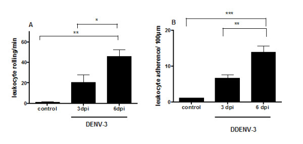
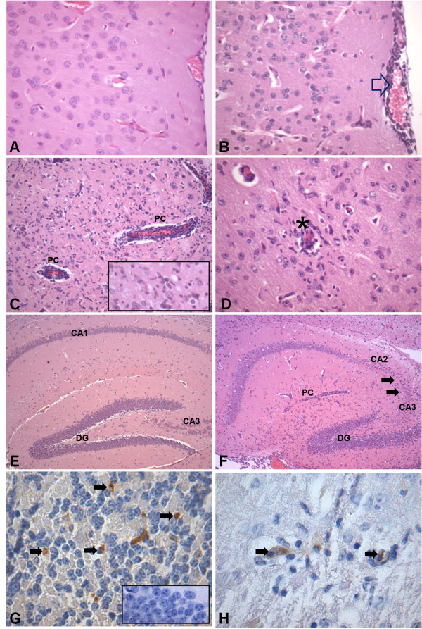
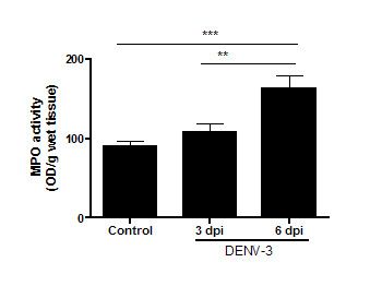
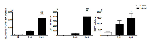
Methods: C57BL/6 mice received 4×10(3) PFU of DENV-3 by an intracranial route. We evaluated the trafficking of leukocytes in brain microvasculature using intravital microscopy, and evaluated chemokine and cytokine profiling by an ELISA test at 3 and 6 days post infection (p.i.). Furthermore, we determined myeloperoxidase activity and immune cell populations, and also performed histopathological analysis and immunostaining for the virus in brain tissue.

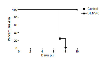
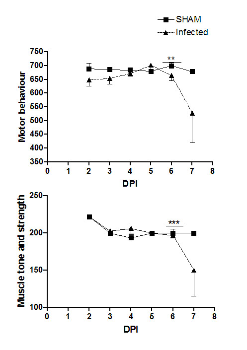
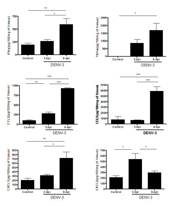
Results: All animals developed signs of encephalitis and died by day 8 p.i. Motor behavior and muscle tone and strength parameters declined at day 7 p.i. We observed increased leukocyte rolling and adhesion in brain microvasculature of infected mice at days 3 and 6 p.i. The infection was followed by significant increases in IFN-γ, TNF-α, CCL2, CCL5, CXCL1, and CXCL2. Histological analysis showed evidence of meningoencephalitis and reactive gliosis. Increased numbers of neutrophils, CD4+ and CD8+ T cells were detected in brain of infected animals, notably at day 6 p.i. Cells immunoreactive for anti-NS-3 were visualized throughout the brain.

Conclusion: Intracerebral infection with non-adapted DENV-3 induces encephalitis and behavioral changes that precede lethality in mice.


Figures

Figure 1
Figure 1
Time course of survival of mice infected with 4 × 10³ PFU of DENV-3 by the intracranial route (n = 20). Control animals (n = 20) received 20 μL of phosphate-buffered saline (PBS).
Figure 2
Figure 2
Visualization of performance (SHIRPA test) of control animals (n = 8) and mice infected with 4 × 10³ PFU of DENV-3 by the intracranial route (n = 8), from days 2 to 7 of infection. Motor behavior (A) and muscle tone and strength parameters (B) showed significant alterations in the infected group at day 7 p.i. ***p < 0.001 and **p < 0.01.
Figure 3
Figure 3
Leukocyte-endothelium interactions visualized using intravital microscopy. Rolling (A) and adhesion (B) of leukocytes in brain microvasculature were assessed in control mice and in mice infected with 4 × 10³ PFU of DENV-3 at 3 and 6 days p.i. by the intracranial route. A higher number of rolling and adhering leukocytes was observed in the microvasculature in brain of infected mice. Data expressed as mean ± SEM of cells per minute (A) and per 100 μm (B). ***p < 0.001, **p < 0.01, *p < 0.05.
Figure 4
Figure 4
Histopathological changes in brain of control mice (n = 4) and of mice infected with 4 × 10³ PFU of DENV-3 (n = 4) by intracranial route, 6 days p.i. Hematoxilin & eosin-stained sections from cerebrum of a control animal with normal histological appearance of cerebrum (A) and hippocampus (E). Brain sections from a DENV-3-infected animal showing: meningoencephalitis characterized by infiltration of immune cells in the meninges (open arrow) (B), perivascular cuffing (PC) (C), gliosis (insert) and (D) vasculitis (asterisk). Note intense neuronal destruction in the pyramidal layer of the CA3 and CA2 areas of the hippocampus (F). Immunohistochemical staining for anti-NS3 protein for DENV-3 showing immunoreactive cells (arrows) in the cerebellar granular layer (G) and in the inflammatory area of cerebrum (H). The insert in G is a negative control from the cerebellum of a non-infected animal. Original magnification: A-B, C (insert), D: x400; C: x200; E-F: x100; G (insert)-H, x1000.
Figure 5
Figure 5
Pathology scores for the brain after DENV-3 infection (C). Morphological analysis was performed on coded samples of different brain areas (cerebrum, cerebellum, hippocampus, brainstem and meninges) at 3 and 6 days p.i. These show progressive pathological changes after DENV-3 infection. Horizontal bars represent median scores.
Figure 6
Figure 6
DENV-3 infection induces cytokines (IFN-γ and TNF-α) and chemokines (CCL2, CCL5, CXCL1, CXCL2) in brain of control mice and of mice infected by the intracranial route with 4 × 10³ PFU of DENV-3 (n = 9, for each time point). Infected mice exhibit high levels of cytokines and chemokines during infection when compared with control animals. Data expressed as mean ± SEM. ***p < 0.001, **p < 0.01, *p < 0.05.
Figure 7
Figure 7
MPO levels in cerebral tissue of control mice and mice infected by the intracranial route with 4 × 10³ PFU of DENV-3. Infected mice showed increased NAG levels on day 6, when compared with the control mice. Data represent results from groups of at least five mice, and results are expressed as mean ± SEM. ***p < 0.001; **p < 0.01.
Figure 8
Figure 8
Brain-sequestered cell numbers in C57Bl/6 mice of control mice (n = 6) and of mice infected with DENV-3. C57Bl/6 mice were infected with 4 × 10³ PFU of DENV-3 by the intracranial route, and assessed at days 3 (n = 4) and 6 p.i. (n = 4). Brain-sequestered cells were counted and then stained with specific antibodies. Flow cytometry, with assessment according to size and granularity, was performed as analysis. Numbers of neutrophils (A), and CD4+ (B) and CD8+ (C) T lymphocytes were evaluated in WT mice. Results are expressed as mean ± SEM, and *p < 0.05, ***p < 0.001 when compared to non-infected mice. ###p < 0.001 when compared to 3 p.i. infected mice.

References

    1. World Health Organization. Impact of dengue. 2009. http://www.who.int/csr/disease/dengue/en/
    1. Figueiredo BL, Cecílio AB, Portela FG, Drumond PB, Oliveira GJ, Bonjardim CA, Ferreira PC, Kroon EG. Dengue virus 3 genotype 1 associated with dengue fever and dengue hemorrhagic fever, Brazil. Emerg Infect Dis. 2008;14:314–316. doi: 10.3201/eid1402.070278. - DOI - PMC - PubMed
    1. Figueiredo RMP, Naveca FG, Bastos MS, Melo MN, Viana SS, Mourão MPG, Costa CA, Gubler IPF. Dengue Virus Type 4, Manaus, Brazil. Emerg Infect Dis. 1998;14:667–669. doi: 10.3201/eid1404.071185. - DOI - PMC - PubMed
    1. Gubler DJ. Dengue and dengue hemorrhagic fever. Clin Microbiol Rev. 1998;11:480–496. - PMC - PubMed
    1. Domingues RB, Kuster GW, Onuki-Castro FL, Souza VA, Levi JE, Pannuti CS. Involvement of the central nervous system in patients with dengue virus infection. J Neurol Sci. 2008;267:36–40. doi: 10.1016/j.jns.2007.09.040. - DOI - PubMed

Publication types