. 2011 Mar 9:8:11.
doi: 10.1186/1743-8977-8-11.

Environmentally persistent free radicals induce airway hyperresponsiveness in neonatal rat lungs

Affiliations

Environmentally persistent free radicals induce airway hyperresponsiveness in neonatal rat lungs

Shrilatha Balakrishna et al. Part Fibre Toxicol. .

Abstract

Background: Increased asthma risk/exacerbation in children and infants is associated with exposure to elevated levels of ultrafine particulate matter (PM). The presence of a newly realized class of pollutants, environmentally persistent free radicals (EPFRs), in PM from combustion sources suggests a potentially unrecognized risk factor for the development and/or exacerbation of asthma.

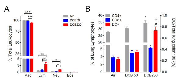
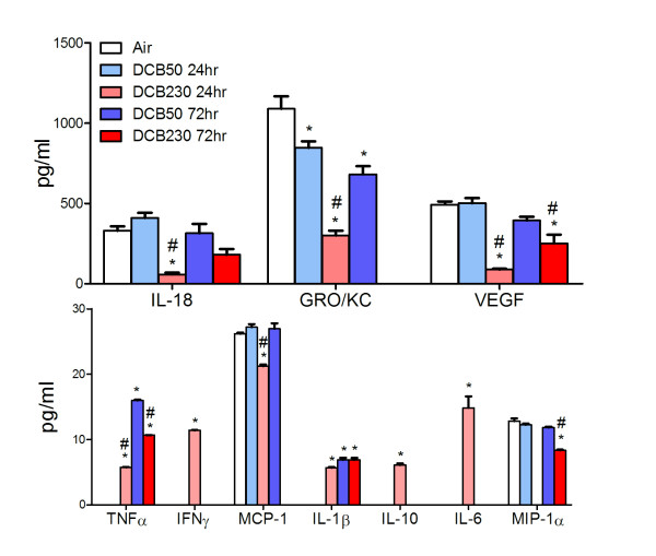
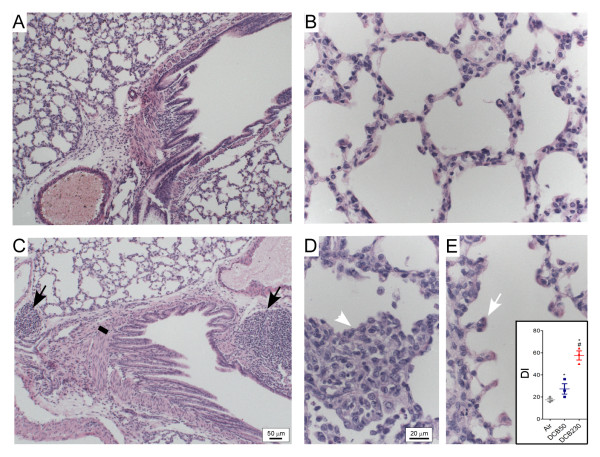
Methods: Neonatal rats (7-days of age) were exposed to EPFR-containing combustion generated ultrafine particles (CGUFP), non-EPFR containing CGUFP, or air for 20 minutes per day for one week. Pulmonary function was assessed in exposed rats and age matched controls. Lavage fluid was isolated and assayed for cellularity and cytokines and in vivo indicators of oxidative stress. Pulmonary histopathology and characterization of differential protein expression in lung homogenates was also performed.

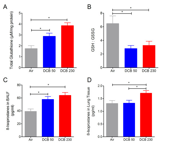
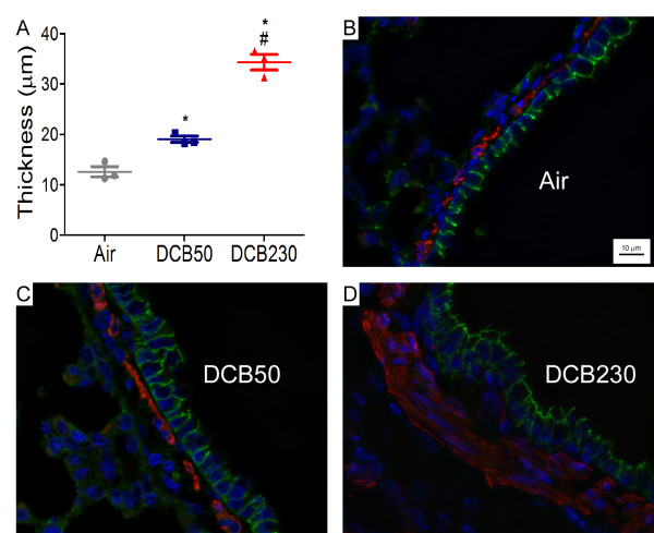
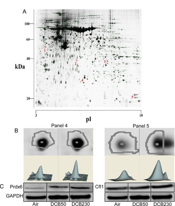
Results: Neonates exposed to EPFR-containing CGUFP developed significant pulmonary inflammation, and airway hyperreactivity. This correlated with increased levels of oxidative stress in the lungs. Using differential two-dimensional electrophoresis, we identified 16 differentially expressed proteins between control and CGUFP exposed groups. In the rats exposed to EPFR-containing CGUFP; peroxiredoxin-6, cofilin1, and annexin A8 were upregulated.

Conclusions: Exposure of neonates to EPFR-containing CGUFP induced pulmonary oxidative stress and lung dysfunction. This correlated with alterations in the expression of various proteins associated with the response to oxidative stress and the regulation of glucocorticoid receptor translocation in T lymphocytes.


Figures

Figure 1
Figure 1
Measurement of airway function following exposure to CGUFP. Neonatal rats (7 d of age) were exposed to CGUFP for 20 m/d for 7 d and pulmonary function was assayed 24 hr after the final exposure. Significant increases in lung resistance (A) in response to MeCh challenge and decreases in compliance (B) among DCB230 exposed neonates compared with control groups (air and DCB50). Quasi-static inflation/deflation curves (i.e. pressure-volume loops) (C) and the area within the curves representative of hysteresis (D). Data are means ± SEM and is representative of three independent experiments with 6 animals/group. *p < 0.05, **p < 0.01, ***p < 0.001.
Figure 2
Figure 2
Indicators of antioxidant status and oxidative stress in postnatal rat lungs following exposure to CGUFP. Changes in total glutathione (A) and GSH:GSSG ratios (B) in whole lung homogenates and 8-Isoprostanes in BALF (C) and whole lung homogenates (D) after CGUFP exposure. All measurements were made 24 hr post-exposure. n = 4-6 animals/group. Data are means ± SEM. *p < 0.05.
Figure 3
Figure 3
Effects of CGUFP exposure on pulmonary inflammation. (A) BALFs were collected and cell differentials obtained. *p < 0.05, **p < 0.01, and ***p < 0.001. (B) Lymphocyte populations in the lungs of rats exposed to CGUFP were quantified by flow cytometry using antibodies specific for the indicated cells after gating on lymphocytes (CD4 and CD8) or non-lymphocytes (DC). Mac indicates macrophages; Neu: neutrophils; Lym: lymphocytes; Eos: eosinophils. n = 4-6 animals/group. *p < 0.05 vs. air and #p < 0.05 vs DCB50.
Figure 4
Figure 4
BALF cytokine levels in postnatal rats after CGUFP exposure. Cytokine levels were determined in the BALF supernatant at 24 hr and 72 hr post-final exposure. n = 6-12 animals/group. Data are expressed as means ± SEM. *p < 0.05 vs Air and #p < 0.05 vs day-matched DCB50.
Figure 5
Figure 5
Light micrographs of exposed rat lungs. Light micrographs of terminal bronchioles (A, C) and alveolar parenchyma (B, D, E) from 15 d old rat lungs following exposure to DCB50, which was visually identical to air (A, B), and DCB230 (C-E). Black arrows denote significant peribronchiolar BALT; line denotes smooth muscle mass surrounding bronchiole (quantified in Figure 6); white arrow denotes lesions of increased alveolar space (quantified in inset of E); and white arrowhead demonstrates alveolar occlusion. Bar represents 50 μm (A, C) and 20 μm (B, D, E).
Figure 6
Figure 6
Quantitation of peribronchiolar smooth muscle content in exposed rat lungs. (A) Quantitative assessment of the thickness of the smooth muscle layer in major airways of 15 d old rat lungs after exposure to air, DCB50 or DCB230. Data represent means ± SEM. n = 3 animals/group. *p < 0.05 vs Air and #p < 0.05 vs DCB50. (B, C, D) Representative micrographs demonstrating expression of α-smooth muscle actin (red) and E-cadherin (green) from 15 d old rat lungs exposed to air, DCB50, or DCB230, respectively. Cell nuclei stained with DAPI (blue).
Figure 7
Figure 7
Proteomic analysis of DCB230 exposed neonatal lungs. (A) 2-D gel proteomic map with selected spots indicating a significant intensity change vs air exposed controls. (B) Enlargement of panels 4 and 5 from air and DCB230 exposed lungs. Panel 4 indicates PRDX6 expression (1.33 increase, p < 0.00034) and panel 5 indicates CFL1 expression (1.34 increase, p < 0.0008). (C) Western blot confirmation of increased expression of both proteins following exposure to DCB230 as compared to air or DCB50. n = 4-6 animals/group.

Similar articles

Cited by

References

    1. Boezen M, Schouten J, Rijcken B, Vonk J, Gerritsen J, van der Zee S, Hoek G, Brunekreef B, Postma D. Peak expiratory flow variability, bronchial responsiveness, and susceptibility to ambient air pollution in adults. Am J Respir Crit Care Med. 1998;158:1848–1854. - PubMed
    1. Pope CA III, Burnett RT, Thun MJ, Calle EE, Krewski D, Ito K, Thurston GD. Lung Cancer, Cardiopulmonary Mortality, and Long-term Exposure to Fine Particulate Air Pollution. JAMA. 2002;287:1132–1141. doi: 10.1001/jama.287.9.1132. - DOI - PMC - PubMed
    1. Schwartz J, Dockery DW, Neas LM, Wypij D, Ware JH, Spengler JD, Koutrakis P, Speizer FE, Ferris BG Jr. Acute effects of summer air pollution on respiratory symptom reporting in children. Am J Respir Crit Care Med. 1994;150:1234–1242. - PubMed
    1. Timonen KL, Pekkanen J. Air pollution and respiratory health among children with asthmatic or cough symptoms. Am J Respir Crit Care Med. 1997;156:546–552. - PubMed
    1. Vedal S, Petkau J, White R, Blair J. Acute effects of ambient inhalable particles in asthmatic and nonasthmatic children. Am J Respir Crit Care Med. 1998;157:1034–1043. - PubMed

Publication types

MeSH terms

LinkOut - more resources