. 2011 May 13;286(19):17181-92.
doi: 10.1074/jbc.M110.179028. Epub 2011 Mar 9.

Solution structures of Ca2+-CIB1 and Mg2+-CIB1 and their interactions with the platelet integrin alphaIIb cytoplasmic domain

Affiliations

Solution structures of Ca2+-CIB1 and Mg2+-CIB1 and their interactions with the platelet integrin alphaIIb cytoplasmic domain

Hao Huang et al. J Biol Chem. .

Abstract

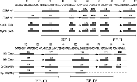
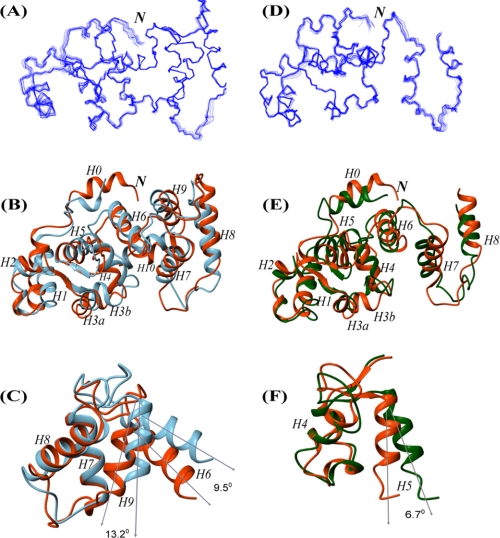
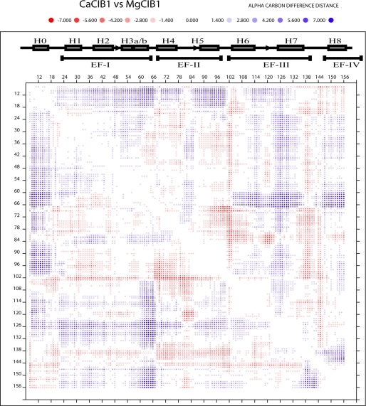
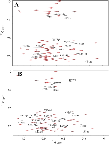
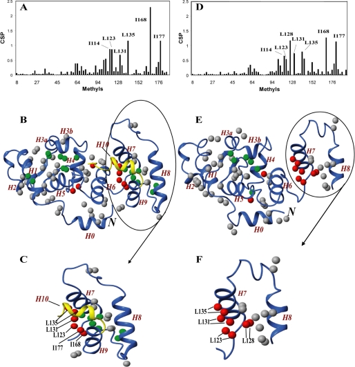
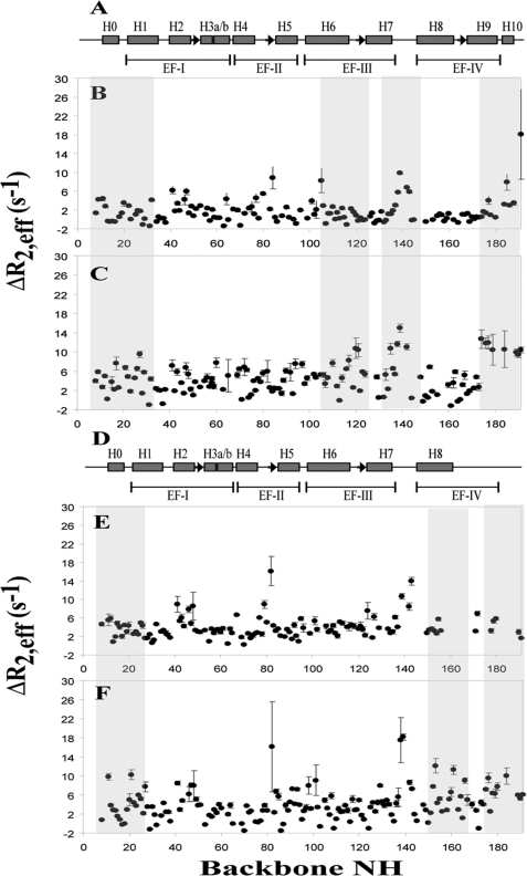
The calcium- and integrin-binding protein 1 (CIB1) is a ubiquitous Ca(2+)-binding protein and a specific binding partner for the platelet integrin αIIb cytoplasmic domain, which confers the key role of CIB1 in hemostasis. CIB1 is also known to be involved in apoptosis, embryogenesis, and the DNA damage response. In this study, the solution structures of both Ca(2+)-CIB1 and Mg(2+)-CIB1 were determined using solution-state NMR spectroscopy. The methyl groups of Ile, Leu, and Val were selectively protonated to compensate for the loss of protons due to deuteration. The solution structure of Ca(2+)-CIB1 possesses smaller opened EF-hands in its C-domain compared with available crystal structures. Ca(2+)-CIB1 and Mg(2+)-CIB1 have similar structures, but the N-lobe of Mg(2+)-CIB1 is slightly more opened than that of Ca(2+)-CIB1. Additional NMR experiments, such as chemical shift perturbation and methyl group solvent accessibility as measured by a nitroxide surface probe, were carried out to further characterize the structures of Ca(2+)-CIB1 and Mg(2+)-CIB1 as well as their interactions with the integrin αIIb cytoplasmic domain. NMR measurements of backbone amide proton slow motion (microsecond to millisecond) dynamics confirmed that the C-terminal helix of Ca(2+)-CIB1 is displaced upon αIIb binding. The EF-hand III of both Ca(2+)-CIB1 and Mg(2+)-CIB1 was identified to be directly involved in the interaction of CIB1 with αIIb. Together, these data illustrate that CIB1 behaves quite differently from related EF-hand regulatory calcium-binding proteins, such as calmodulin or neuronal calcium sensor proteins.


Figures

FIGURE 1.
FIGURE 1.
Secondary structure arrangements of the two available x-ray crystal structures of Ca2+-CIB1 (1XO5 and 1Y1A) and the NMR solution structures of Ca2+-CIB1 and Mg2+-CIB1. The positions of the four EF-hand helix-loop-helix structures are indicated. Boxes indicate α-helices, and arrows indicate β-strands.
FIGURE 2.
FIGURE 2.
Correlation between the Ca2+-CIB1 and Mg2+-CIB1 solution structures revealed by RDC analysis. The backbone NH-RDC of Mg2+-CIB1 shows a good correlation with the solution structure of Ca2+-CIB1 (A), in which the outliers are labeled. The absolute values of the difference between the observed RDC and calculated RDC are plotted as a function of the primary sequence (B), and a horizontal line highlights the outliers.
FIGURE 3.
FIGURE 3.
Solution structures of Ca2+-CIB1 and Mg2+-CIB1. A, ensemble of the 20 best solution structures of Ca2+-CIB1. B, superimposed solution structure (red) and crystal structure (1XO5, blue) of Ca2+-CIB1 with an r.m.s.d. of 2.71 Å for the well defined areas (residues 24–136 and 146–191). C, superimposed EF-III (H6 and H7) and EF-IV (H8 and H9) region (residues 102–180) of the Ca2+-CIB1 crystal (1XO5, blue) and solution structures (red) demonstrate that the solution structure has a more closed conformation than the crystal structure. The two structures were superimposed based on H7 and H8; thus, the opening was highlighted by the orientations of H6 and H9, respectively. D, ensemble of the calculated 20 best structures of Mg2+-CIB1. E, superimposed solution structure of Ca2+-CIB1 (red) and Mg2+-CIB1 (green) with an r.m.s.d. of 3.27 Å for the well defined areas (residues 24–136 and 146–157). F, superimposed EF-II (H4 and H5) of the solution structures of Ca2+-CIB1 (red) and Mg2+-CIB1 (green) demonstrate that Mg2+-CIB1 has a more opened conformation than Ca2+-CIB1. The two structures were superimposed based on H4; thus, the opening was highlighted by the orientations of H5. The letter N in italics indicates the N terminus of the protein.
FIGURE 4.
FIGURE 4.
Difference distance matrix plot to compare the structural differences between Ca2+-CIB1 and Mg2+-CIB1 (residues 8–157). The secondary structure was labeled with filled boxes indicating helices and arrows indicating β-strands. The program DDMP gives real values of differences concerning Cα atoms in two structures. For example, the distance between the Cα atoms of i and j residues in the Ca2+-CIB1 structure is 10 Å, and the corresponding distance in structure Mg2+-CIB1 is 15 Å, and hence [(Ca2+-CIB1)-(Mg2+-CIB1)]ij = 10–15 = −5 Å. The red and blue color shading is indicated above the figure.
FIGURE 5.
FIGURE 5.
Superimposed 13C-HSQC spectra of the Ile, Leu, and Val methyl groups of CIB1 and CIB1 bound to αIIb in the absence (black) and in the presence of 6 eq of TEMPOL (red). The same amount of TEMPOL (6 eq) was added into 12C,2H,15N-uniformly and Ile-δ1-13CH3,Leu,Val-13CH3,12CD3-labeled Ca2+-CIB1 (A) and Mg2+-CIB1 (B) samples at the same concentration (0.3 mm). The peaks marked with an asterisk are most likely from the 13C isotopic natural abundance of TEMPOL as those peaks do not appear for the protein alone.
FIGURE 6.
FIGURE 6.
Using Ile-δ1,Leu-δ1,δ2,Val-γ1,γ2-methyl groups to probe the interaction between CIB1 and αIIb. The methyl CSP of CIB1 upon binding of αIIb is plotted as a function of methyl residues for Ca2+-CIB1 (A) and Mg2+-CIB1 (D). Methyl groups were mapped on the solution structure of Ca2+-CIB1 (B) and Mg2+-CIB1 (E) (residues 8–157). All methyl groups were labeled as round spheres, with nonaffected or marginally affected (CSP <0.2) methyl groups in gray, slightly affected (0.2 ≤ CSP <0.5) methyl groups in green, and significantly affected (CSP >0.5) methyl groups in red. The C-terminal extension (residues 179–191) of the Ca2+-CIB1 solution structure is colored golden to demonstrate the C-terminal displacement mechanism. C and F show the significantly affected methyl groups for Ca2+- and Mg2+-CIB1 upon binding of the αIIb peptide, respectively. The letter N in italics indicates the N terminus of the protein.
FIGURE 7.
FIGURE 7.
Slow motion dynamics (microsecond to millisecond) backbone CPMG experiments of both CIB1 and CIB1 complexed with αIIb. A, secondary structure of Ca2+-CIB1, and microsecond dynamics of Ca2+-CIB1 (B) and Ca2+-CIB1 (C) in complex with αIIb; D, secondary structure of Mg2+-CIB1, and microsecond time scale dynamics of Mg2+-CIB1 (E) and Mg2+-CIB1 (F) in complex with αIIb. These experiments were acquired on a 2H,15N-uniformly labeled CIB1 at 37 °C, pH 7.5, at a field strength of 500 MHz. The errors were obtained based on the analysis of duplicate experiments.

Similar articles

Cited by

References

    1. Gifford J. L., Walsh M. P., Vogel H. J. (2007) Biochem. J. 405, 199–221 - PubMed
    1. Yamniuk A. P., Vogel H. J. (2006) Calcium Bind. Proteins 1, 150–155
    1. Leisner T. M., Yuan W., DeNofrio J. C., Liu J., Parise L. V. (2007) Curr. Opin. Hematol. 14, 255–261 - PubMed
    1. Naik U. P., Patel P. M., Parise L. V. (1997) J. Biol. Chem. 272, 4651–4654 - PubMed
    1. Jarman K. E., Moretti P. A., Zebol J. R., Pitson S. M. (2010) J. Biol. Chem. 285, 483–492 - PMC - PubMed

Publication types