. 2011 Apr 29;286(17):14762-9.
doi: 10.1074/jbc.M110.185181. Epub 2011 Mar 9.

Syk regulates multiple signaling pathways leading to CX3CL1 chemotaxis in macrophages

Affiliations

Syk regulates multiple signaling pathways leading to CX3CL1 chemotaxis in macrophages

Haein Park et al. J Biol Chem. .

Abstract

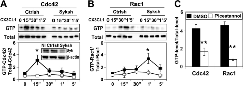
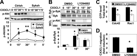
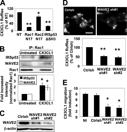
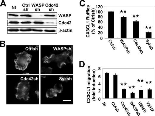
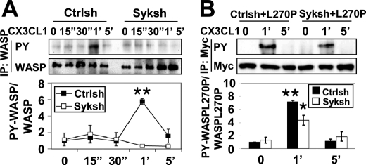
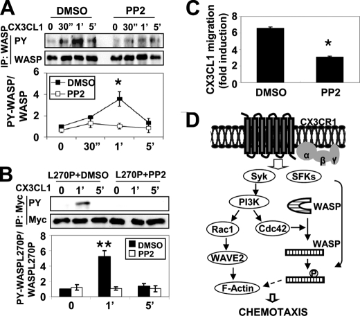
Several studies have clearly established the importance of the interaction between macrophages and CX3CL1 in the progression of disease. A previous study demonstrated that Syk was required for CX3CL1-mediated actin polymerization and chemotaxis. Here, we delineated the signaling cascade of Syk-mediated cell migration in response to CX3CL1. Inhibition of Syk in bone marrow-derived macrophages or reduction of Syk expression using siRNA in RAW/LR5 cells indicated that Syk was required for the activation of PI3K, Cdc42, and Rac1. Also, reduction in WASP or WAVE2 levels, common downstream effectors of Cdc42 or Rac1, resulted in impaired cell migration to CX3CL1. Syk indirectly regulated WASP tyrosine phosphorylation through Cdc42 activation. Altogether, our data identify that Syk mediated chemotaxis toward CX3CL1 by regulating both Rac1/WAVE2 and Cdc42/WASP pathways, whereas Src family kinases were required for proper WASP tyrosine phosphorylation.


Figures

FIGURE 1.
FIGURE 1.
CX3CL1-induced Rho GTPases activation requires Syk. Cells were incubated with 50 ng/ml CX3CL1 for the indicated times, and Cdc42 and Rac1 activity was determined as described under “Experimental Procedures.” A, representative blot and quantification of GTP-bound Cdc42 normalized to total Cdc42 (Total) relative to control shRNA-treated (Ctrlsh) cells at time 0 are shown. The inset shows a representative blot of noninfected (NI) and puromycin-resistant stable populations of Ctrlsh or Syk shRNA-treated cells (Syksh) immunoblotted for Syk and β-actin. B, a representative blot and quantification of Rac1 activity relative to Ctrlsh cells at time 0 are shown (n = 3). Data represent mean ± S.E. *, p < 0.05 compared with Ctrlsh cells at time 0. C, BMMs pretreated with DMSO (vehicle) or 50 μm piceatannol were stimulated with CX3CL1 and Cdc42 activity at 15 s (15″) or Rac1 activity at 1 min (1′) was determined (n = 3). Data represent mean ± S.E. **, p < 0.01 compared with DMSO-treated BMMs.
FIGURE 2.
FIGURE 2.
PI3K activation by Syk is required for CX3CL1-induced Rho GTPase activation and chemotaxis. A, Ctrlsh and Syk shRNA-treated cells (Syksh) were stimulated with CX3CL1 for the indicated times, and the activation of Akt was assessed by Akt phosphorylation (p-Akt). Representative blots of p-Akt and total Akt are shown. p-Akt levels were quantified by densitometry and normalized to total Akt. Akt activity relative to Ctrlsh cells at time 0 was plotted (n = 3). Data represent mean ± S.E. *, p < 0.05 and **, p < 0.01 compared with Ctrlsh cells at time 0. B, RAW/LR5 cells were preincubated for 30 min with DMSO (vehicle) or 100 μm LY294002 prior to CX3CL1 stimulation for the indicated times. Representative blot of immunoprecipitated Syk (IP) probed for either phospho-specific Syk or Syk (upper panels). LY294002 efficacy was monitored by p-Akt levels in whole cell lysates (WCL; middle panels). Quantification of Syk activity relative to DMSO-treated cells at time 0 is shown (lower panels). C, BMMs pre-treated with DMSO (vehicle) or 100 μm LY294002 were incubated with CX3CL1 for the indicated times, and Cdc42 or Rac1 activity was determined at 15 sec (15″) or 1 min (1′), respectively, as described in the legend to Fig. 1 (n = 3). Mean ± S.E. *, p < 0.05 compared with DMSO-treated BMMs. D, RAW/LR5 cells were preincubated for 30 min with DMSO (vehicle) or 100 μm LY294002 before being subjected to a Transwell migration assay. CX3CL1-induced chemotaxis was expressed as fold induction compared with DMSO-treated cells in the absence of CX3CL1 (n = 3). Mean ± S.E. *, p < 0.05 compared with DMSO-treated cells.
FIGURE 3.
FIGURE 3.
WAVE2 activation by Rac1 through IRSp53 is required for ruffle formation and cell migration in response to CX3CL1. A, RAW/LR5 cells expressing dominant-negative Myc-tagged Rac1 (Rac1N17), Myc-tagged Rac2 (Rac2N17), or HA-tagged IRSp53 deficient in WAVE2 binding (IRSp53ΔSH3) were stimulated with CX3CL1 for 1 min. Cells expressing the constructs were identified by epitope staining (Myc for Rac1/2 and HA for IRSp53) and F-actin-rich protrusions (ruffles) were visualized by Alexa Fluor 568 phalloidin staining. The number of CX3CL1-elicited protrusions (CX3CL1 ruffles) was quantified as described under “Experimental Procedures” and expressed as a percentage of the CX3CL1 stimulation observed in nontransfected cells (NT) on the same coverslip (n = 3). Mean ± S.E. *, p < 0.05 and **, p < 0.01 compared with nontransfected cells. B, RAW/LR5 cells were either left untreated or subjected to CX3CL1 stimulation for 1 min prior to lysis and immunoprecipitation (IP) of Rac1 followed by Western blotting with the indicated antibodies. A representative blot is shown with quantification of IRSp53 (black bar) or WAVE2 (white bar) co-immunoprecipitated by Rac (n = 3). Mean ± S.E. *, p < 0.05 compared with untreated cells. C, a representative Western blot of WAVE2 and β-actin expression in RAW/LR5 cells infected with either control sequence (Ctrlsh) or two different shRNA sequences targeting WAVE2 (sh#1 and sh#2). D, representative images of Ctrlsh or WAVE2 shRNA-treated cells stimulated with CX3CL1 for 1 min and stained for F-actin with Alexa Fluor 568 phalloidin staining (upper panels). The number of CX3CL1-elicited protrusions was quantified as described in A and expressed as a percentage of the CX3CL1 stimulation observed in Ctrlsh-treated cells (lower panel) (n = 3). Mean ± S.E. **, p < 0.01 compared with Ctrlsh-treated cells. Scale bar = 10 μm. E, CX3CL1 chemotaxis of the indicated cell lines was determined using a Transwell assay and expressed as fold induction compared with the corresponding condition in the absence of CX3CL1 (n = 3). Mean ± S.E. *, p < 0.05 compared with noninfected (NI) cells.
FIGURE 4.
FIGURE 4.
Cdc42 binding and tyrosine phosphorylation of WASP are required for CX3CL1-induced cell migration. A, RAW/LR5 cells were infected with a retrovirus bearing a control sequence (Ctrlsh) or a sequence against WASP (WASPsh) or Cdc42 (Cdc42sh). A representative blot of lysates from noninfected (NI) and puromycin-resistant stable populations of infected cells immunoblotted against WASP, Cdc42, and β-actin are shown (n = 5). B, Ctrlsh, WASPsh, Cdc42sh, and Syk shRNA-treated (Syksh) cells were stimulated with CX3CL1 for 1 min, and ruffles were visualized by Alexa Fluor 568 phalloidin staining. Representative images of three independent experiments are shown. Scale bar, 10 μm. C, the number of CX3CL1-elicited protrusions (CX3CL1 ruffles) was expressed as a percentage of the CX3CL1 stimulation observed in Ctrlsh cells (n = 3). Mean ± S.E. **, p < 0.01 compared with Ctrlsh cells. D, chemotactic ability of the indicated cells was determined using a Transwell assay. In addition, clones of WASPsh cells expressing human Myc-tagged WASP deficient in Cdc42 binding (H246D) or phosphorylation-deficient WASP (Y291F; Ref. 20) were tested. Data were expressed as fold induction compared with the corresponding condition in the absence of CX3CL1 (n = 3). Mean ± S.E. **, p < 0.01 compared with CX3CL1-induced migration in noninfected (NI) cells.
FIGURE 5.
FIGURE 5.
Syk regulates tyrosine phosphorylation of WASP through Cdc42 activation. A, control (Ctrlsh) and Syk shRNA-treated cells (Syksh) were stimulated with CX3CL1 for the indicated times. WASP was immunoprecipitated (IP) using WASP antibody followed by Western blotting for phosphotyrosine (PY) and WASP. Blots were quantified by densitometry and normalized to total WASP. A representative blot and quantification of WASP tyrosine phosphorylation relative to Ctrlsh cells at time 0 are shown (n = 3). Mean ± S.E. **, p < 0.01 compared with Ctrlsh cells at time 0. B, Ctrlsh or Syk shRNA-treated cells were transiently transfected with Myc-tagged constitutively active WASP (L270P) and then stimulated with CX3CL1 for the indicated times. Myc-tagged L270P WASP was immunoprecipitated using Myc antibody following by Western blotting for phosphotyrosine and Myc. Blots were quantified by densitometry and normalized to Myc. A representative blot and quantification of L270P WASP tyrosine phosphorylation relative to Ctrlsh cells at time 0 are shown (n = 3). Mean ± S.E. *, p < 0.05 and **, p < 0.01 compared with Ctrlsh cells at time 0. 15″, 15 sec; 1′, 1 min.
FIGURE 6.
FIGURE 6.
SFKs regulate WASP tyrosine phosphorylation. A, RAW/LR5 cells were pretreated for 1 h with DMSO (vehicle) or 10 μm PP2 prior to CX3CL1 stimulation for the indicated times. WASP was immunoprecipitated (IP) followed by Western blotting for phosphotyrosine (PY) and WASP. Blots were quantified by densitometry and normalized to amount of WASP. A representative blot and quantification of WASP tyrosine phosphorylation relative to DMSO-treated cells at time 0 are shown (n = 3). Mean ± S.E. *, p < 0.05 compared with DMSO-treated cells at time 0. B, RAW/LR5 cells expressing Myc-L270P WASP were preincubated with DMSO (vehicle) or 10 μm PP2 prior to CX3CL1 stimulation for the indicated times. Myc-tagged L270P WASP was immunoprecipitated using Myc antibody followed by Western blotting for phosphotyrosine and Myc. Blots were quantified by densitometry and normalized to Myc. A representative blot and quantification of L270P WASP tyrosine phosphorylation relative to DMSO-treated cells at time 0 are shown (n = 3). Mean ± S.E. **, p < 0.01 compared with DMSO-treated cells at time 0. C, RAW/LR5 cells were preincubated for 1 h with DMSO (vehicle) or 10 μm PP2 before being subjected to a Transwell migration assay. CX3CL1-induced chemotaxis was expressed as fold induction compared with DMSO-treated cells in the absence of CX3CL1 (n = 3). Mean ± S.E. *, p < 0.05 compared with DMSO-treated cells. D, model of Syk-mediated signal pathways in response to CX3CL1. Syk and SFKs are activated independently by CX3CL1, leading to efficient chemotaxis. Syk regulates both Cdc42/WASP and Rac1/WAVE2 pathways through activation of PI3K. Syk is required to activate Cdc42, which induces a conformational change in WASP permitting phosphorylation by SFKs (see “Discussion” for details). 30″, 30 sec; 1′, 1 min.

Similar articles

Cited by

References

    1. Cambien B., Pomeranz M., Schmid-Antomarchi H., Millet M. A., Breittmayer V., Rossi B., Schmid-Alliana A. (2001) Blood 97, 2031–2037 - PubMed
    1. Umehara H., Bloom E. T., Okazaki T., Nagano Y., Yoshie O., Imai T. (2004) Arterioscler. Thromb. Vasc. Biol. 24, 34–40 - PubMed
    1. Imai T., Hieshima K., Haskell C., Baba M., Nagira M., Nishimura M., Kakizaki M., Takagi S., Nomiyama H., Schall T. J., Yoshie O. (1997) Cell 91, 521–530 - PubMed
    1. Goda S., Imai T., Yoshie O., Yoneda O., Inoue H., Nagano Y., Okazaki T., Imai H., Bloom E. T., Domae N., Umehara H. (2000) J. Immunol. 164, 4313–4320 - PubMed
    1. Ruth J. H., Volin M. V., Haines G. K., 3rd, Woodruff D. C., Katschke K. J., Jr., Woods J. M., Park C. C., Morel J. C., Koch A. E. (2001) Arthritis Rheum. 44, 1568–1581 - PubMed

Publication types

MeSH terms