. 2011 Apr 22;286(16):14659-69.
doi: 10.1074/jbc.M110.183269. Epub 2011 Mar 9.

Roles of chromatin remodeling factors in the formation and maintenance of heterochromatin structure

Affiliations

Roles of chromatin remodeling factors in the formation and maintenance of heterochromatin structure

Qun Yu et al. J Biol Chem. .

Abstract

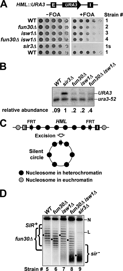
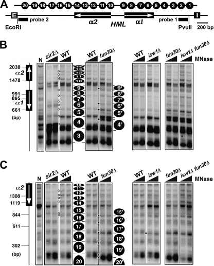
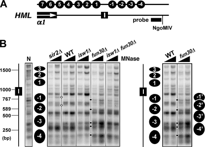
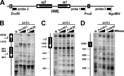
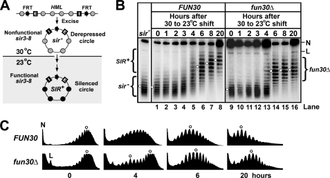
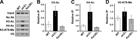
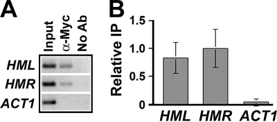
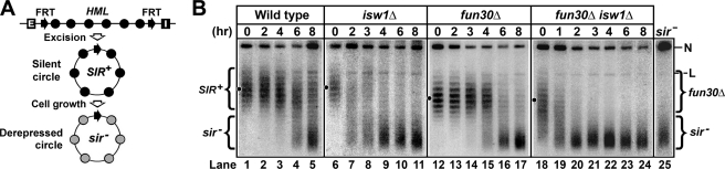
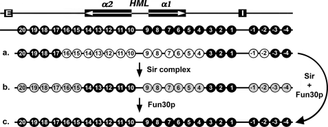
Heterochromatin consists of highly ordered nucleosomes with characteristic histone modifications. There is evidence implicating chromatin remodeling proteins in heterochromatin formation, but their exact roles are not clear. We demonstrate in Saccharomyces cerevisiae that the Fun30p and Isw1p chromatin remodeling factors are similarly required for transcriptional silencing at the HML locus, but they differentially contribute to the structure and stability of HML heterochromatin. In the absence of Fun30p, only a partially silenced structure is established at HML. Such a structure resembles fully silenced heterochromatin in histone modifications but differs markedly from both fully silenced and derepressed chromatin structures regarding nucleosome arrangement. This structure likely represents an intermediate state of heterochromatin that can be converted by Fun30p to the mature state. Moreover, Fun30p removal reduces the rate of de novo establishment of heterochromatin, suggesting that Fun30p assists the silencing machinery in forming heterochromatin. We also find evidence suggesting that Fun30p functions together with, or after, the action of the silencing machinery. On the other hand, Isw1p is dispensable for the formation of heterochromatin structure but is instead critically required for maintaining its stability. Therefore, chromatin remodeling proteins may rearrange nucleosomes during the formation of heterochromatin or serve to stabilize/maintain heterochromatin structure.


Figures

FIGURE 1.
FIGURE 1.
Effects of fun30Δ and isw1Δ on transcriptional silencing and DNA supercoiling at HML. A, FUN30 and ISW1 are required for efficient HML silencing. Top, a schematic of the HML locus bearing a URA3 marker gene in strains 1–4 and 1s is shown. HML-E and -I silencers are shown as filled boxes with white letters. Bottom, growth phenotypes of strains 1–4 and 1s are shown. Cells of each strain were grown to log phase, and serial 10-fold dilutions were spotted and grown on synthetic medium with (+) or without (−) 1 mg/ml of FOA. B, Northern blot analysis of URA3 expression is shown. Total RNA was extracted from log phase cells of each strain, and 10 μg was loaded in each lane. URA3 mRNA was detected by Northern blotting and hybridization with a radioactive probe made from the coding sequence of URA3. Note that in the strains tested here, the transcript from the nonfunctional ura3-52 allele at the normal URA3 locus on chromosome V is truncated, due to the insertion of a Ty element into the URA3 coding sequence. Therefore, transcripts from both URA3 gene at HML and ura3-52 allele could be examined simultaneously. As the normal URA3 locus is not subject to SIR-dependent silencing, ura3-52 mRNA was used as an internal control for loading. The intensity of each band was quantified using NIH image, and the relative abundance of URA3 mRNA in each strain was calculated as the ratio of URA3 mRNA signal over ura3-52 mRNA signal. C, shown is the strategy for examining the topology of HML DNA (27). Two FRT (Flp1p recombination target) sequences (filled arrows) are inserted to flank HML. Recombination between FRTs by the site-specific recombinase Flp1p excises HML as a circular minichromosome. D, FUN30 but not ISW1 is required for the high negative supercoiling of HML DNA. DNA isolated from strains 5 (WT), 6 (fun30Δ), 7 (isw1Δ), 8 (fun30Δ isw1Δ), and 9 (sir3Δ) was subjected to agarose gel electrophoresis in the presence of 26 μg/ml chloroquine. After Southern blotting, topoisomers of the HML circle were detected by an HML-specific probe. Under the conditions employed here, more negatively supercoiled topoisomers migrate more slowly. The center of distribution of topoisomers from each strain is indicated by a dot. The nicked/relaxed and linear forms of the HML circle are indicated as N and L, respectively. Topoisomers of HML circle from WT, fun30Δ and sir3Δ strains are collectively designated SIR+, fun30Δ, and sir, respectively.
FIGURE 2.
FIGURE 2.
Effects of fun30Δ and isw1Δ on the primary chromatin structure of HML. A, shown is the HMLα locus. The HML-E and -I silencers and the α1 and α2 genes are shown. Filled bars indicate the sequences of probes 1 and 2 used in indirect end labeling experiments shown in B and C, respectively. The positions of 20 nucleosomes inferred from a previous mapping experiment (11) are shown at the top. B and C, examination of HML chromatin in strains 10 (WT), 11 (sir2Δ), 12 (fun30Δ), 13 (isw1Δ), and 14 (fun30Δ isw1Δ) by MNase digestion and indirect end labeling is shown. DNA isolated from MNase-treated chromatin was digested with PvuII (B) or EcoRI (C) and fractionated on agarose gels. Genomic (naked) DNA from strain 10 (WT) was also treated with MNase and then digested with PvuII (B) or EcoRI (C). This sample is designated N. After Southern-blotting, DNA fragments were detected by probe 1 near the PvuII site (B) or probe 2 near the EcoRI site (C). The relative positions of the α1 and α2 genes are shown on the left. The positions of inferred nucleosomes are indicated by filled ovals. Some bands representing MNase sensitive sites are indicated by open diamonds or (see “Results” for descriptions).
FIGURE 3.
FIGURE 3.
Effects of fun30Δ and isw1Δ on the primary chromatin structure around the HML-I silencer. A, schematics of the region around HML-I are shown. The filled bar indicates the sequence of probe 3 used in the indirect end labeling experiment shown in B. The positions of nucleosomes 1–7 mapped previously (11) and -1 through -4 mapped in B are indicated at the top. B, mapping chromatin around HML-I in strains 10–14 is shown. DNA isolated from MNase treated chromatin was digested with NgoMIV and fractionated on an agarose gel. Genomic DNA (naked DNA, designated N) from strain 10 was also treated with MNase and then digested with NgoMIV. After Southern blotting, DNA fragments ending at the NgoMIV site were detected by hybridization with probe 3. The position of the HML-I silencer is shown on the left. The positions of inferred nucleosomes are indicated by filled ovals. Some bands representing MNase-sensitive sites are indicated by open diamonds or black dots (see “Results” for descriptions). Note the WT and fun30Δ lanes were aligned side by side on the right for better comparison and illustration of the positions of inferred nucleosomes.
FIGURE 4.
FIGURE 4.
Effects of fun30Δ on the primary chromatin structure of HML in the sir background. A, shown is the HMLα locus. Filled bars indicate the sequences of probes 1–3 used in indirect end labeling experiments shown in B–D, respectively. B–D, examination of HML chromatin in strains 11 (sir2Δ) and 15 (sir2Δ fun30Δ) by MNase digestion and indirect end labeling. MNase-treated chromatin was digested with PvuII (B), EcoRI (C), or NgoMIV (D) and fractionated on an agarose gel. After Southern blotting, DNA fragments ending at the PvuII (B), EcoRI (C), or NgoMIV (D) site were detected by hybridization with probes 1–3, respectively. The position of the α genes and HML-I silencer are shown on the left of the blots. N, naked DNA.
FIGURE 5.
FIGURE 5.
Effect of fun30Δ on de novo establishment of HML heterochromatin structure. A, shown is a strategy for examining the de novo establishment of heterochromatin on circular HML minichromosome. Shaded and filled circles denote nucleosomes in derepressed chromatin and heterochromatin, respectively. The HML locus including the E and I silencers is flanked by a pair of FRTs. See “Results” for description of the scheme. B, examination of the kinetics of establishment of heterochromatin on HML circle in strains 16 (sir3-8 FUN30) and 17 (sir3-8 fun30Δ). Cells of each strain grown at 30 °C in YPR medium were treated with galactose for 2.5 h to induce excision of the HML circle. Cells were then shifted to fresh YPD (yeast extract/peptone + glucose) medium and incubated at 23 °C for up to 20 h. Aliquots of the culture were harvested at time points 0, 1, 2, 3, 4, 6, 8, and 20 h. DNA was isolated and fractionated by agarose gel electrophoresis in the presence of 26 μg/ml chloroquine. Under the conditions used, more negatively supercoiled topoisomers run more slowly. N and L, nicked and linear forms of the HML circle, respectively. The topoisomers corresponding to the silent and derepressed states of HML circles are designated SIR+ and sir, respectively. C, the profiles of topoisomers in lanes 1, 5, 6, and 8 and 9, 13, 14, and 16 were determined using the NIH image software. The centers of distribution of topoisomers are marked by open circles.
FIGURE 6.
FIGURE 6.
Deletion of FUN30 does not affect histone hypoacetylation and hypomethylation in heterochromatin at HML. A, the abundance of an HML sequence in strain 10 (WT), 11 (sir2Δ), or 12 (fun30Δ) was measured by PCR before (Input) and after chromatin IP with antibodies for acetylated histones H3 and H4 (H3-Ac and H4-Ac), and histone H3 methylated at K79 (H3-K79-Me) as well as total H3 (H3). No Ab, samples from mock ChIP without using antibody. The gel picture of PCR products from one of three independent experiments is shown. The data were quantified and plotted in B–D. The value of relative IP of H3-Ac, H4-Ac, or H3-K79-Me was calculated as the ratio of signal (IP/input) for H3-Ac (B), H4-Ac (C), or H3-K79-Me (D) over that for total H3. The means of data from all three independent experiments together with corresponding S.D. are presented. The value for each WT sample is taken as 1.
FIGURE 7.
FIGURE 7.
Fun30 is enriched at HML and HMR loci. A, the abundance of HML, HMR, and ACT1 sequences in strain 18 carrying FUN30-Myc was measured by PCR before (Input) and after (α-Myc) chromatin IP with α-Myc antibody. No Ab, samples from mock ChIP without using antibody. The gel picture of PCR products from one of three independent experiments is shown. The data were quantified and are plotted in B. The value of relative IP was calculated as the ratio of IP signal over input signal. The means of data from all three independent experiments together with corresponding S.D. are presented. The value for HMR sequence is taken as 1.
FIGURE 8.
FIGURE 8.
ISW1 is required for maintaining the stability of HML heterochromatin. A, a strategy for examining the stability of heterochromatin on HML minichromosome dissociated from silencers is shown. Filled and shaded circles denote nucleosomes in silent and derepressed chromatins, respectively. See “Results” for a description. B, examination of the kinetics of the conversion of silent HML circle (SIR+) to derepressed circle (sir) in strains 5 (wild type), 6 (fun30Δ), 7 (isw1Δ), and 8 (fun30Δ isw1Δ) is shown. Cells of each strain grown to stationary phase in YPR were treated with galactose for 2.5 h to induce the excision of the HML circle. Cells were then shifted and diluted into fresh yeast extract/peptone/glucose medium and further incubated for 8 h. Aliquots of culture were harvested at time points 0, 1, 2, 3, 4, 6, and 8 h. DNA was isolated from the samples and fractionated by agarose gel electrophoresis in the presence of chloroquine. N and L, nicked and linear forms of the HML circle, respectively. Topoisomers corresponding to the silent and derepressed states of HML circles are designated SIR+ and sir, respectively. Topoisomers of HML circle from the fun30Δ strain are designated fun30Δ.
FIGURE 9.
FIGURE 9.
Contribution of Fun30p to the special primary structure of HML heterochromatin. Illustrated is a summary of chromatin mapping data shown in Figs. 2 and 3. The HML locus and nucleosomes 1–20 and -1 through -4 in silent (SIR+) HML heterochromatin are shown as filled ovals at the top. a, shown is nucleosome distribution in HML chromatin in the derepressed state (in the absence of Sir complex). Open circles represent the nucleosomes in the derepressed (sir) state that differ in position and/or stability from their counterparts in the silent (SIR+) state. b, shown is nucleosome distribution in HML chromatin in an intermediate state (in the absence of Fun30p). Shaded circles represent the nucleosomes in the intermediate (fun30Δ) state that differ in position and/or stability from their counterparts in the silent (SIR+) state. c, shown is nucleosome distribution in HML chromatin in fully silenced heterochromatin state.

Similar articles

Cited by

References

    1. Grewal S. I., Moazed D. (2003) Science 301, 798–7802 - PubMed
    1. Dillon N. (2004) Biol. Cell 96, 631–637 - PubMed
    1. Sun F. L., Cuaycong M. H., Elgin S. C. (2001) Mol. Cell. Biol. 21, 2867–2879 - PMC - PubMed
    1. Wallrath L. L., Elgin S. C. (1995) Genes Dev. 9, 1263–1277 - PubMed
    1. Bassett A. R., Cooper S. E., Ragab A., Travers A. A. (2008) PLoS One 3, e2099. - PMC - PubMed

Publication types

MeSH terms