

Islet-1 regulates Arx transcription during pancreatic islet alpha-cell development
- PMID: 21388963
- PMCID: PMC3083195
- DOI: 10.1074/jbc.M111.231670
Islet-1 regulates Arx transcription during pancreatic islet alpha-cell development
Abstract
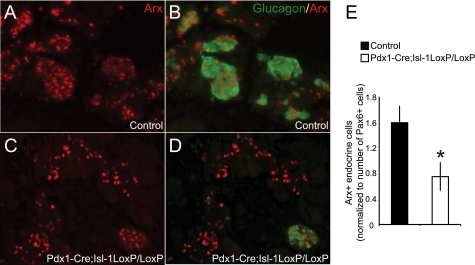
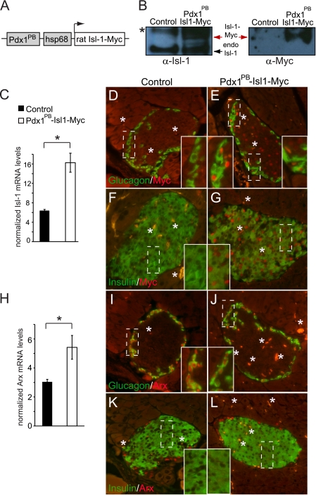
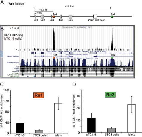
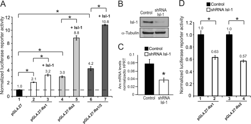
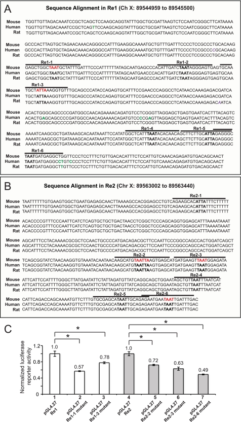
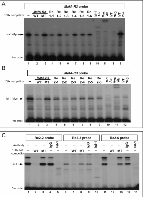
Aristaless related homeodomain protein (Arx) specifies the formation of the pancreatic islet α-cell during development. This cell type produces glucagon, a major counteracting hormone to insulin in regulating glucose homeostasis in adults. However, little is known about the factors that regulate Arx transcription in the pancreas. In this study, we showed that the number of Arx(+) cells was significantly reduced in the pancreata of embryos deficient for the Islet-1 (Isl-1) transcription factor, which was also supported by the reduction in Arx mRNA levels. Chromatin immunoprecipitation analysis localized Isl-1 activator binding sites within two highly conserved noncoding regulatory regions (Re) in the Arx locus, termed Re1 (+5.6 to +6.1 kb) and Re2 (+23.6 to +24 kb). Using cell line-based transfection assays, we demonstrated that a Re1- and Re2-driven reporter was selectively activated in islet α-cells, a process mediated by Isl-1 in overexpression, knockdown, and site-directed mutation experiments. Moreover, Arx mRNA levels were up-regulated in islet α-cells upon Isl-1 overexpression in vivo. Isl-1 represents the first known activator of Arx transcription in α-cells, here established to be acting through the conserved Re1 and Re2 control domains.
Figures
Similar articles
-
Islet α-, β-, and δ-cell development is controlled by the Ldb1 coregulator, acting primarily with the islet-1 transcription factor.Diabetes. 2013 Mar;62(3):875-86. doi: 10.2337/db12-0952. Epub 2012 Nov 27. Diabetes. 2013. PMID: 23193182 Free PMC article.
-
Present status and expectation of aristaless-related homeobox (ARX) in endocrine pancreas.Int J Dev Biol. 2019;63(11-12):579-587. doi: 10.1387/ijdb.190242sx. Int J Dev Biol. 2019. PMID: 32149367 Review.
-
The islet-expressed Lhx1 transcription factor interacts with Islet-1 and contributes to glucose homeostasis.Am J Physiol Endocrinol Metab. 2019 Mar 1;316(3):E397-E409. doi: 10.1152/ajpendo.00235.2018. Epub 2019 Jan 8. Am J Physiol Endocrinol Metab. 2019. PMID: 30620636 Free PMC article.
-
Activation of amylin gene transcription by LIM domain homeobox gene isl-1.Mol Endocrinol. 1996 Mar;10(3):243-51. doi: 10.1210/mend.10.3.8833653. Mol Endocrinol. 1996. PMID: 8833653
-
Transcriptional regulation of α-cell differentiation.Diabetes Obes Metab. 2011 Oct;13 Suppl 1:13-20. doi: 10.1111/j.1463-1326.2011.01440.x. Diabetes Obes Metab. 2011. PMID: 21824252 Review.
Cited by
-
Gene regulatory networks governing pancreas development.Dev Cell. 2013 Apr 15;25(1):5-13. doi: 10.1016/j.devcel.2013.03.016. Dev Cell. 2013. PMID: 23597482 Free PMC article. Review.
-
Integration of ATAC-seq and RNA-seq identifies human alpha cell and beta cell signature genes.Mol Metab. 2016 Jan 11;5(3):233-244. doi: 10.1016/j.molmet.2016.01.002. eCollection 2016 Mar. Mol Metab. 2016. PMID: 26977395 Free PMC article.
-
Alpha TC1 and Beta-TC-6 genomic profiling uncovers both shared and distinct transcriptional regulatory features with their primary islet counterparts.Sci Rep. 2017 Sep 20;7(1):11959. doi: 10.1038/s41598-017-12335-1. Sci Rep. 2017. PMID: 28931935 Free PMC article.
-
Cell type and tissue specific function of islet genes in zebrafish pancreas development.Dev Biol. 2013 Jun 1;378(1):25-37. doi: 10.1016/j.ydbio.2013.03.009. Epub 2013 Mar 19. Dev Biol. 2013. PMID: 23518338 Free PMC article.
-
Elevation of transcription factor Islet-1 levels in vivo increases β-cell function but not β-cell mass.Islets. 2012 May-Jun;4(3):199-206. doi: 10.4161/isl.19982. Epub 2012 May 1. Islets. 2012. PMID: 22595886 Free PMC article.
References
-
- Burcelin R., Katz E. B., Charron M. J. (1996) Diabetes Metab. 22, 373–396 - PubMed
-
- Dobbs R., Sakurai H., Sasaki H., Faloona G., Valverde I., Baetens D., Orci L., Unger R. (1975) Science 187, 544–547 - PubMed
-
- Toft I., Gerich J. E., Jenssen T. (2002) Metabolism 51, 1128–1134 - PubMed
-
- Unger R. H. (1971) Diabetes 20, 834–838 - PubMed
Publication types
MeSH terms
Substances
Grants and funding
LinkOut - more resources
Full Text Sources
Molecular Biology Databases
