

doi: 10.1128/JVI.00239-11.
Epub 2011 Mar 9.
Subclinical brain injury caused by H5N1 influenza virus infection
Affiliations
- PMID: 21389133
- PMCID: PMC3126180
- DOI: 10.1128/JVI.00239-11
Item in Clipboard
Subclinical brain injury caused by H5N1 influenza virus infection
J Virol.
2011 May.
Abstract
Although H5N1 influenza A viruses can cause systemic infection, their neurotropism and long-term effects on the central nervous system (CNS) are not fully understood. We assessed H5N1viral invasion of the CNS and its long-term effects in a ferret model. An H5N1 virus caused nonsuppurative encephalitis, which lasted for 3 months without neurologic signs. Further, another H5N1 virus caused nonsuppurative vasculitis with brain hemorrhage. Three-dimensional analysis of viral distribution in the brain identified the olfactory system as a major route for brain invasion. The efficient growth of virus in the upper respiratory tract may thus facilitate viral brain invasion.
Figures
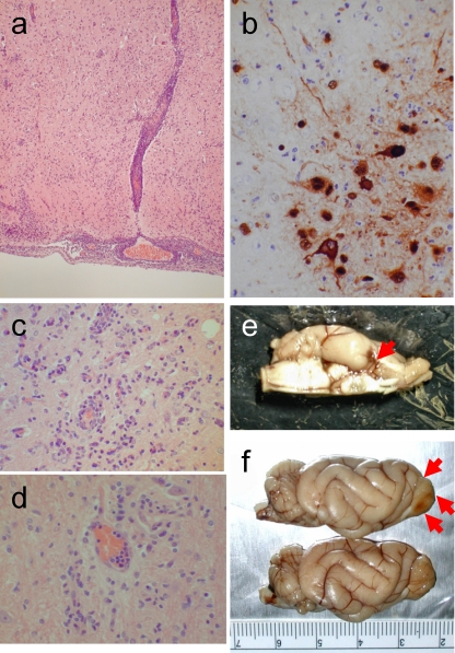
Brain lesions in HK486 virus-infected ferrets. (a) Severe nonsuppurative encephalitis in the olfactory area at day 6 postinfection (p.i.). (b) Viral antigen expression in a brain lesion at day 12 p.i. Neuronal and glial cells are stained with anti-H5 virus antiserum. (Inset) Noninfected neurons and glia. (c) Smoldering encephalitis in brain tissue at 3 months p.i. (d) Perivascular glial scar formation at 9 months p.i. (e) Macroscopically visible brain lesion in part of the olfactory system (piriform lobe) on day 12 p.i. (f) Partial loss of an olfactory bulb (upper side of brain [red arrows]) due to viral encephalitis at 1 month p.i. Compare these images with the brain from an age-matched control (lower brain).
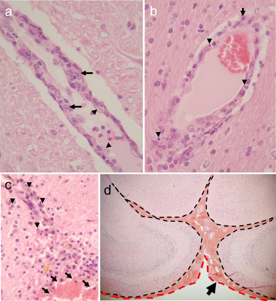
Brain lesions in HK483 virus-infected ferrets. (a) Prominent nonsuppurative vasculitis at day 6 p.i. Note the severe swelling of a vascular endothelial cell (arrowheads) and migration of macrophages into the vascular wall (arrows), compared with the normal appearance of the surrounding brain parenchyma. (b) Scattered apoptotic cells (arrowheads) and polymorphonuclear leukocytes (arrow) in the vascular wall on day 6 p.i. (c) Old and fresh hemorrhagic lesions in the thalamus of a ferret that underwent necropsy at 6 months p.i. (arrowheads, hemosiderin-laden macrophages in an old lesion; arrows, fresh hemorrhage with red blood cells). (d) Fresh subarachnoid hemorrhage in a ferret brain at 6 months p.i. Note the accumulation of red blood cells between leptomeninges (dashed black line) and arachnoid mater (dashed red line).
Distribution of brain lesions following infection with the HK483, HK486, HK213, NCVD18, or VN1204 strain of H5N1 virus. Brain lesions are mapped on three-dimensional images of a yellow mongoose brain. Selected parts of the brain sections were analyzed; therefore, the plots of the lesion locations are discontinuous. (a) Olfactory route (yellow). The distribution of lesions and viral antigens (red) associated with HK213 (b) or NCVD18 (c) infection follows the olfactory route (dashed yellow line). In animals infected with HK486 (d) or VN1204 (e), the lesions and viral antigens are located in the brain stem (white arrows) and the olfactory route (red plots within the dashed yellow line). The HK483 strain (f) caused severe blood vessel damage, with apparent hemorrhagic lesions (blue plots). (g) Posterior view of the olfactory route (yellow plots). (h) Posterior view of HK483-induced hemorrhagic lesions (blue) and vasculitis (red) outside the olfactory route. Panels a to f are ventral views.
References
-
- Abdel-Ghafar A. N., et al. 2008. Update on avian influenza A (H5N1) virus infection in humans. N. Engl. J. Med. 358:261–273 - PubMed
-
- Anderson W. W., Jaros R. M. 1958. Neurologic complications of Asian influenza. Neurology 8:568–570 - PubMed
-
- Bell W. E., McKee A. P., Utterback R. A. 1958. Asian influenza virus as the cause of acute encephalitis. Neurology 8:500–502 - PubMed
-
- de Jong M. D., et al. 2005. Fatal avian influenza A (H5N1) in a child presenting with diarrhea followed by coma. N. Engl. J. Med. 352:686–692 - PubMed
Publication types
MeSH terms
LinkOut - more resources
Full Text Sources
Medical
