. 2011 Mar 9;31(10):3650-61.
doi: 10.1523/JNEUROSCI.5565-10.2011.

Structural and mechanistic determinants of a novel site for noncompetitive inhibition of GluN2D-containing NMDA receptors

Affiliations

Structural and mechanistic determinants of a novel site for noncompetitive inhibition of GluN2D-containing NMDA receptors

Kasper B Hansen et al. J Neurosci. .

Abstract

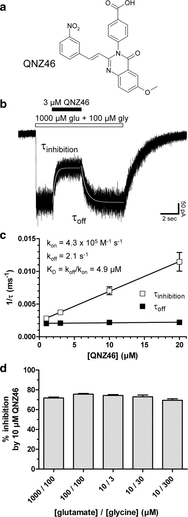
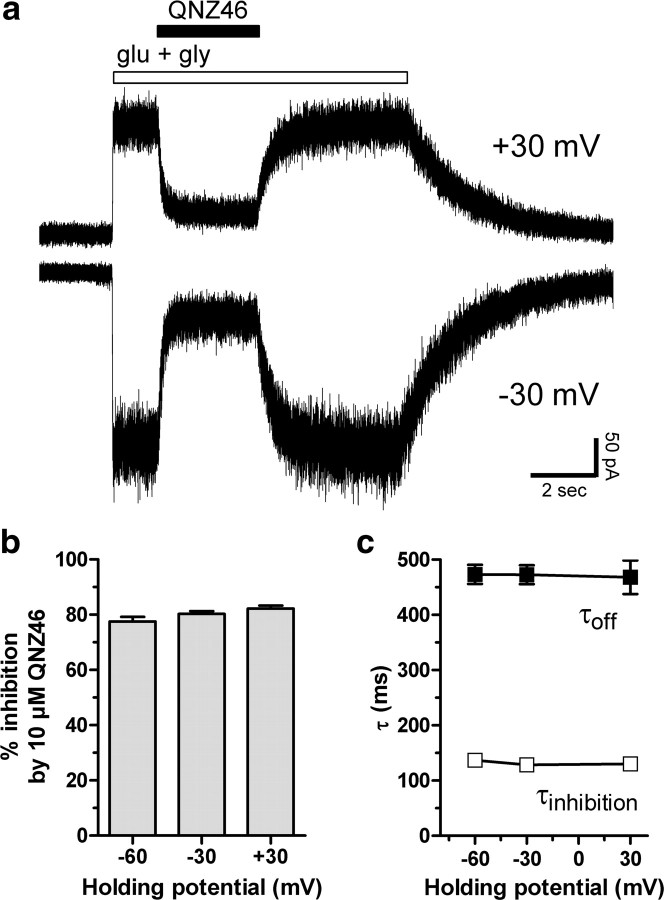
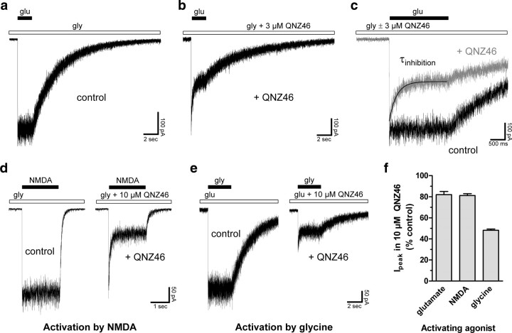
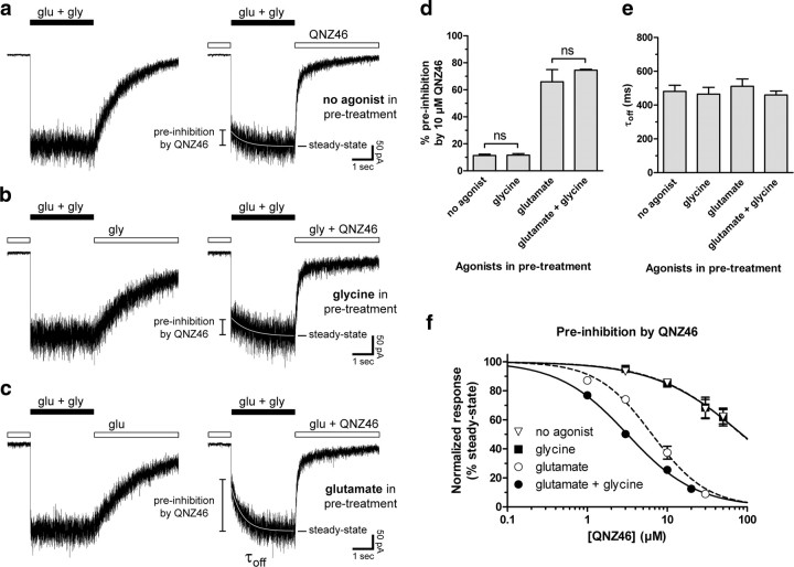
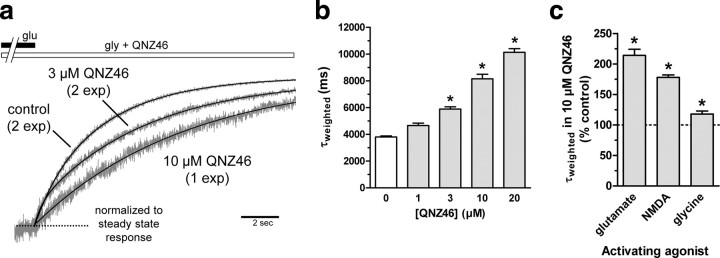
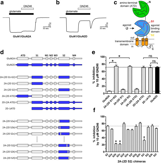
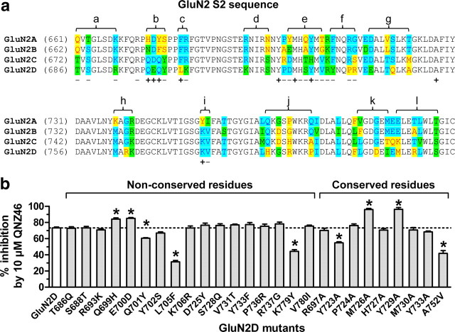
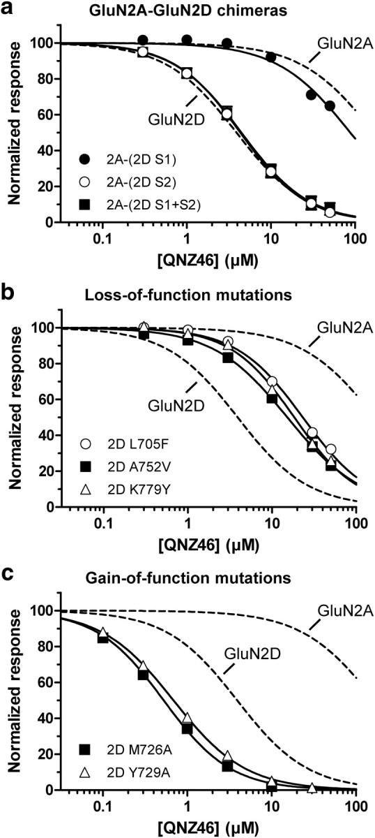
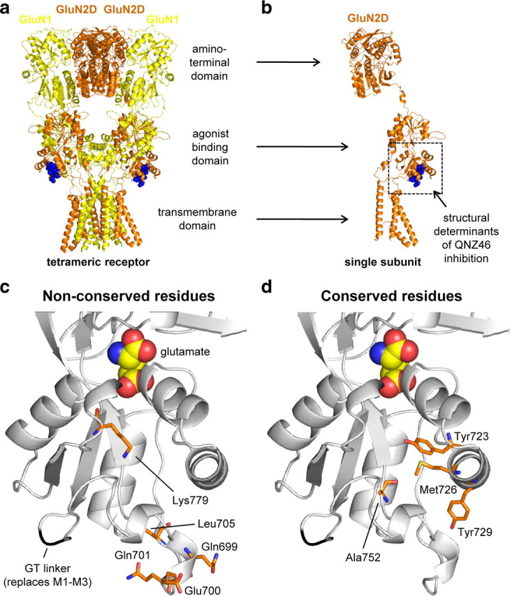
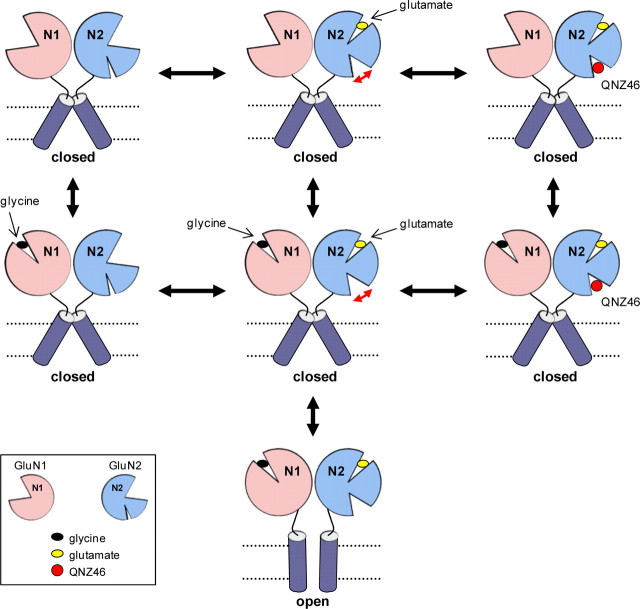
NMDA receptors are ionotropic glutamate receptors that mediate excitatory synaptic transmission and have been implicated in several neurological diseases. We have evaluated the mechanism of action of a class of novel subunit-selective NMDA receptor antagonists, typified by (E)-4-(6-methoxy-2-(3-nitrostyryl)-4-oxoquinazolin-3(4H)-yl)-benzoic acid (QNZ46). We found that QNZ46 inhibits NMDA receptor function in a noncompetitive and voltage-independent manner by an unconventional mechanism that requires binding of glutamate to the GluN2 subunit, but not glycine binding to the GluN1 subunit. This dependency of antagonist association on glutamate binding to GluN2 renders these compounds nominally use-dependent, since inhibition will rely on synaptic release of glutamate. Evaluation of the structural determinants responsible for the subunit-selectivity of QNZ46 revealed that these compounds act at a new site that has not previously been described. Residues residing in the part of the agonist binding domain immediately adjacent to the transmembrane helices appear to control selectivity of QNZ46 for GluN2C- and GluN2D-containing receptors. These residues are well-positioned to sense glutamate binding to GluN2 and thus to mediate glutamate-dependent actions. This new class of noncompetitive antagonists could provide an opportunity for the development of pharmacological tools and therapeutic agents that target NMDA receptors at a new site and modulate function by a novel mechanism.


Figures

Figure 1.
Figure 1.
QNZ46 is a noncompetitive NMDA receptor antagonist. a, Chemical structure of QNZ46. QNZ46 represents a new class of NMDA receptor antagonists and is ∼50-fold selective for GluN2C- and GluN2D-containing receptors over receptors containing GluN2A or GluN2B (Mosley et al., 2010) (see Table 1; supplemental Fig. S1, available at www.jneurosci.org as supplemental material). b, Representative whole-cell current response recorded under voltage-clamp from recombinant GluN1/GluN2D receptors expressed in a HEK293 cell using rapid solution exchange. The onset and offset of QNZ46 inhibition of steady-state responses to saturating glutamate and glycine can be described using monoexponential fits (see Table 2). c, τinhibition and τoff were used to determine the association and dissociation rates of QNZ46 binding as a function of concentration, allowing estimation of KD for antagonist binding to GluN1/GluN2D (N = 4). d, Inhibition of GluN1/GluN2D by QNZ46 was independent of agonist concentration and could not be surmounted by increasing concentrations of glutamate or glycine (values from 3 to 11 cells were not significantly different; p > 0.05; one-way ANOVA with Tukey–Kramer post-test). This is consistent with a noncompetitive mechanism of action (see also supplemental Fig. S1, available at www.jneurosci.org as supplemental material).
Figure 2.
Figure 2.
Inhibition by QNZ46 is voltage-independent. a, Representative whole-cell current responses recorded under voltage-clamp at +30 and −30 mV from recombinant GluN1/GluN2D receptors. 10 μm QNZ46 and 1000 μm glutamate plus 100 μm glycine (glu + gly) were applied to the cell for the duration of the respective bars above the recorded traces. b, Inhibition by 10 μm QNZ46 is independent of membrane holding potential (values from 5 to 7 cells were not significantly different; p > 0.05; one-way ANOVA with Tukey–Kramer post-test). c, Time constants for inhibition (τinhibition) and recovery from inhibition (τoff) by 10 μm QNZ46 at GluN1/GluN2D are independent of membrane holding potential (values from 5 to 7 cells were not significantly different; p > 0.05; one-way ANOVA with Tukey–Kramer post-test). Error bars are SEM, shown when larger than symbol.
Figure 3.
Figure 3.
Inhibition by QNZ46 is use-dependent. a, b, Representative whole-cell current responses from recombinant GluN1/GluN2D receptors. Glutamate (1000 μm; glu) was applied to the cell using rapid solution exchange in the continuous presence of 100 μm glycine alone (gly; control) or in continuous presence of 100 μm glycine plus 3 μm QNZ46 (+ QNZ46). Rapid application of glutamate in the continuous presence of QNZ46 induces a rapid response that peaks and then relaxes to a steady-state level. c, Overlay of the traces in a and b. d, Representative whole-cell current responses activated by NMDA from recombinant GluN1/GluN2D receptors. NMDA (200 μm) was applied to the cell using rapid solution exchange in the continuous presence of 100 μm glycine alone (gly; control) or in continuous presence of 100 μm glycine plus 10 μm QNZ46 (+ QNZ46). Similar to activation by glutamate, activation by the partial glutamate site agonist NMDA in the continuous presence of QNZ46 induces a rapid response that peaks and then relaxes to a steady-state level. e, Representative whole-cell current responses activated by glycine from recombinant GluN1/GluN2D receptors. Glycine (300 μm) was applied to the cell using rapid solution exchange in the continuous presence of 10 μm glutamate alone (glu; control) or in continuous presence of 10 μm glutamate plus 10 μm QNZ46 (+ QNZ46). Activation by glycine in the continuous presence of QNZ46 and glutamate leads to responses that show considerable inhibition of the instantaneous current, evidenced by the small peak current in the presence QNZ46 compared with control. f, Summary of peak current responses (Ipeak) in the continuous presence of QNZ46 relative to control peak current responses in the absence of QNZ46. The activating agonist is indicated; when glutamate and NMDA were activating agonists, the cells were bathed in glycine, and when glycine was activating agonist, the cells were bathed in glutamate (N = 5–8) (see also supplemental Table 4, available at www.jneurosci.org as supplemental material).
Figure 4.
Figure 4.
Inhibition by QNZ46 depends on glutamate binding. a–c, Representative whole-cell current responses from recombinant GluN1/GluN2D receptors. a, Responses from a cell that experienced a solution exchange from ligand-free extracellular solution (left) or QNZ46 (10 μm) alone (right) to glutamate (1000 μm) plus glycine (300 μm). b, Responses from a cell that experienced a solution exchange from glycine (100 μm) (left) or glycine plus QNZ46 (10 μm) (right) to glutamate (1000 μm) plus glycine (100 μm). c, Responses from a cell that experienced a solution exchange from glutamate (10 μm) (left) or glutamate plus QNZ46 (10 μm) (right) to glutamate (10 μm) plus glycine (300 μm). d, Preinhibition of GluN1/GluN2D pretreated with QNZ46 alone (no agonist; as in a) or glycine plus QNZ46 (glycine; as in b) were significantly different from preinhibition of receptors pretreated with glutamate plus QNZ46 (glutamate; as in c) or glutamate plus glycine plus QNZ46 (glutamate + glycine; as in Fig. 1b) (N = 4–7; p < 0.05; one-way ANOVA with Tukey–Kramer post-test; see supplemental Table 4, available at www.jneurosci.org as supplemental material). e, The time constants (τoff) for the slow relaxation to full amplitude steady-state following receptor washout of 10 μm QNZ46 are independent of whether glycine, glutamate, or no agonist was present before receptor activation (values from 4 to 7 cells were not significantly different; p > 0.05; one-way ANOVA with Tukey–Kramer post-test; see supplemental Table 4, available at www.jneurosci.org as supplemental material). f, Concentration-response data for preinhibition by QNZ46 represented as the instantaneous current normalized to the full amplitude steady-state responses (i.e., 100% corresponds to no preinhibition). The IC50 values for QNZ46 preinhibition were 3.2 μm (N = 4) when both glutamate and glycine were present during pretreatment (see also supplemental Fig. S2, available at www.jneurosci.org as supplemental material) and 6.4 μm (N = 5) when glutamate alone was present during pretreatment. In the presence of glycine alone or no agonists, the IC50 values were 85 μm (N = 5) or 86 μm (N = 5), respectively.
Figure 5.
Figure 5.
QNZ46 prolongs receptor deactivation time. a, Overlay of whole-cell current recordings as shown in Figure 3b normalized to steady-state responses to glutamate plus glycine in the absence (control) or presence of 3 or 10 μm QNZ46. The deactivation time courses in the absence of QNZ46 and in the presence of 3 μm QNZ46 were best described using dual-exponential fits (2 exp), whereas the deactivation time course in the presence of 10 μm QNZ46 was best described using a monoexponential fit (1 exp). b, Analyses of the deactivation time course following removal of glutamate in the continuous presence of QNZ46 show that the time constants for deactivation (τweighted) significantly increase up to approximately 2-fold in the presence of QNZ46 (1–20 μm) (see supplemental Table 2, available at www.jneurosci.org as supplemental material). *Significantly different from τweighted in the absence of QNZ46 (white bar) (p < 0.05; one-way ANOVA with Tukey–Kramer post-test). Data are from four to seven cells. c, QNZ46 prolongs deactivation of responses to both glutamate and the partial agonist NMDA in the continuous presence of glycine. The QNZ46-mediated prolongation of deactivation glycine responses in the continuous presence of glutamate is less pronounced compared with the effects on glutamate and NMDA deactivation (see also supplemental Table 4, available at www.jneurosci.org as supplemental material). *Significantly different from control in the absence of QNZ46 (indicated by the dashed line; p < 0.05; two-tailed unpaired t test). Data are from five to seven cells.
Figure 6.
Figure 6.
Recovery from QNZ46 inhibition is not use-dependent. a, Representative overlay of whole-cell current recordings from one HEK293 cell expressing recombinant GluN1/GluN2D receptors. The cell is initially stepped into NMDA (200 μm) plus glycine (300 μm), and the resulting response is then inhibited by QNZ46 (10 μm). Subsequent to this inhibition, the cell is stepped into wash (i.e., no agonists and no QNZ46) at time 0 (t = 0). The cell is then stepped back into NMDA plus glycine at different time intervals (Δt). QNZ46 unbinding and recovery from inhibition occur from the step into wash (t = 0) to the time point where the steady-state response is reached following the step back into NMDA plus glycine. NMDA was used as glutamate site agonist because the time constant for NMDA deactivation (τweighted 92 ms; see supplemental Table 4, available at www.jneurosci.org as supplemental material) is 41-fold lower (i.e.faster) than τ weighted for glutamate (3810 ms; see supplemental Table 2, available at www.jneurosci.org as supplemental material), and 3.5-fold lower than τoff for QNZ46 in the continuous presence of NMDA (320 ms). By contrast, τweighted for glutamate is 8.3-fold higher (i.e., slower) than τoff for QNZ46 in the continuous presence of glutamate (460 ms; see supplemental Table 2, available at www.jneurosci.org as supplemental material). b, Representative recordings showing recovery from inhibition on an expanded time scale (a, boxed area). c, Mean recovery from inhibition at different intervals in wash (no agonist) measured as the instantaneous current response relative to the steady-state response from the same recording are shown as white circles with the monoexponential fit (black line) (τoff = 290 ± 10; N = 6). The averaged time course for recovery from inhibition in the continuous presence of NMDA from the same cells is shown as gray dots (with agonist) (τoff = 320 ± 20; N = 6). The time constants for recovery from QNZ46 inhibition in the presence and absence of agonist are not significantly different (p > 0.05, two-tailed unpaired t test).
Figure 7.
Figure 7.
Structural determinants for subunit-selective QNZ46 inhibition are located in the S2 segment of the agonist binding domain. a, b, Representative two-electrode voltage-clamp recordings from GluN1/GluN2A (a) and GluN1/GluN2D (b) receptors expressed in Xenopus oocytes. The receptors were activated by glutamate (100 μm) and inhibited by QNZ46 (10 μm). Glycine (30 μm) was present in all solutions. c, NMDA receptors are comprised of four semiautonomous domains; the extracellular amino-terminal domain (ATD) and agonist binding domain, the transmembrane domain containing three transmembrane helices (M1, M2, and M4) and a membrane re-entrant loop (M2), and the intracellular C-terminal domain. The agonist binding domain is formed by two amino acid segments (S1 and S2). d, Linear representations of the polypeptide chains of GluN2A (gray) and GluN2D (blue), as well as chimeric GluN2A-GluN2D subunits (for chimeric junctions, see supplemental Table 1, available at www.jneurosci.org as supplemental material). e, Summary of the inhibition of the response to glutamate (100 μm) by 10 μm QNZ46 in the continuous presence of glycine (30 μm) (as shown in a, b) for five chimeric GluN2 subunits and a deletion mutant of GluN2D without the amino-terminal domain (all GluN2 subunits coexpressed with GluN1 in Xenopus oocytes). *Significantly different from GluN1/GluN2A (white bar) (p < 0.05; one-way ANOVA with Tukey–Kramer post-test). ns, Not significantly different from GluN1/GluN2D (black bar) (p > 0.05; one-way ANOVA with Tukey–Kramer post-test). Data are from 4 to 13 oocytes. f, The QNZ46-sensitivity of 2A-(2D S2) chimeras (coexpressed with GluN1) that progressively dissect the S2 region identified three distinct regions that significantly influence QNZ46 selectivity (see also Fig. 8a). *Significantly different from 2A-(2D S2) (S2; white bar) (p < 0.05; one-way ANOVA with Tukey–Kramer post-test). Data are from 4 to 12 oocytes.
Figure 8.
Figure 8.
Individual residues that influence subunit-selective QNZ46 inhibition. a, Amino acid sequence alignment of the S2 segment from GluN2A-D. The regions marked above the sequences were converted to the GluN2A sequence in the 2A-(2D S2) chimera of the indicated letter. For example, Thr686, Ser688, and Arg693 were converted to Gln, Thr, and Lys, respectively, in the 2A-(2D S2a) chimera. −, the residue was mutated in GluN2D with no effect on QNZ46 sensitivity; +, QNZ46 sensitivity was significantly changed when the residue in GluN2D was mutated. b, Individual nonconserved residues in the three regions identified by the 2A-(2D S2b), 2A-(2D S2c), and 2A-(2D S2i) chimeras (see Fig. 7f), as well as surrounding residues were mutated in GluN2D to the corresponding residues in GluN2A. Furthermore, selected residues in GluN2D that are conserved in GluN2A and are located close to Gln699, Glu700, Gln701, Leu705, and Lys779 in a homology model of GluN2D (see Fig. 10) were mutated to alanine (or valine in case of Ala752). Nine mutations significantly changed the sensitivity to QNZ46. *Significantly different from GluN1/GluN2D (white bar) (p < 0.05; one-way ANOVA with Tukey–Kramer post-test). Data are from 4 to 20 oocytes.
Figure 9.
Figure 9.
Individual residues that influence QNZ46 inhibition. ac, Concentration-response data for QNZ46 inhibition of GluN2A-GluN2D chimeras (a), GluN2D loss-of-function mutants (b), and GluN2D gain-of-function mutants (coexpressed with GluN1) (c) (for IC50 values, see Table 4). For comparison, the concentration-response curves for QNZ46 at GluN1/GluN2A and GluN1/GluN2D are shown as dashed lines only (data points are included in supplemental Fig. S1, available at www.jneurosci.org as supplemental material).
Figure 10.
Figure 10.
Mapping of residues that influence QNZ46 inhibition. a, Illustration of residues important for QNZ46 inhibition in a hypothetical tetrameric NMDA receptor structure using the crystal structure of the tetrameric membrane-spanning GluA2 as guide (Protein Data Bank ID 3KG2) (Sobolevsky et al., 2009). The GluN1 (yellow) and GluN2D (orange) subunits are colored according to the position they would occupy in a tetrameric NMDA receptor (Sobolevsky et al., 2009). Residues in GluA2 homologous to residues in GluN2D that influence QNZ46 activity are highlighted as blue spheres. b, Same as in a, but here only a single subunit is shown. c, Close-up of the lower portion of the agonist binding domain proximal to the membrane from a homology model of the isolated GluN2D agonist binding domain with bound glutamate (see Materials and Methods). Bound glutamate in the agonist binding pocket is shown as spheres. The main chain of the Gly-Thr (GT) linker that replaces the transmembrane helices M1–M3 is shown in black (Furukawa et al., 2005). The five residues identified in this study that influence QNZ46 inhibition and are nonconserved between GluN2A and GluN2D are highlighted as orange sticks. d, The four identified residues that influence QNZ46 inhibition and are conserved between GluN2A and GluN2D are highlighted as orange sticks.
Figure 11.
Figure 11.
Working hypothesis for noncompetitive and glutamate-dependent inhibition of NMDA receptors by QNZ46. Glutamate binding promotes cleft closure of the GluN2 agonist binding domain, and a conformational change within the GluN2 subunit either increases the accessibility of the QNZ46 binding site or places the binding site in a higher affinity conformation (indicated by red arrow). Occupation of the QNZ46 binding site then inhibits opening of the gate. The glutamate-dependent enhancement of QNZ46 binding accounts for the apparent use-dependence of QNZ46 inhibition, since the receptor can enter the open state before QNZ46 can bind. Glycine binding to the GluN1 subunit alone without occupation at the glutamate binding site in GluN2 does not promote rearrangement of GluN2 in a manner that enhances QNZ46 binding.

References

    1. Balannik V, Menniti FS, Paternain AV, Lerma J, Stern-Bach Y. Molecular mechanism of AMPA receptor noncompetitive antagonism. Neuron. 2005;48:279–288. - PubMed
    1. Costa BM, Irvine MW, Fang G, Eaves RJ, Mayo-Martin MB, Skifter DA, Jane DE, Monaghan DT. A novel family of negative and positive allosteric modulators of NMDA receptors. J Pharmacol Exp Ther. 2010;335:614–621. - PMC - PubMed
    1. Eswar N, Eramian D, Webb B, Shen MY, Sali A. Protein structure modeling with MODELLER. Methods Mol Biol. 2008;426:145–159. - PubMed
    1. Furukawa H, Singh SK, Mancusso R, Gouaux E. Subunit arrangement and function in NMDA receptors. Nature. 2005;438:185–192. - PubMed
    1. Horak M, Vlcek K, Chodounska H, Vyklicky L., Jr Subtype-dependence of N-methyl-D-aspartate receptor modulation by pregnenolone sulfate. Neuroscience. 2006;137:93–102. - PubMed

Publication types

Substances

LinkOut - more resources