. 2011 Mar 9;31(10):3805-12.
doi: 10.1523/JNEUROSCI.5561-10.2011.

Distinct neural substrates of duration-based and beat-based auditory timing

Affiliations

Distinct neural substrates of duration-based and beat-based auditory timing

Sundeep Teki et al. J Neurosci. .

Abstract

Research on interval timing strongly implicates the cerebellum and the basal ganglia as part of the timing network of the brain. Here we tested the hypothesis that the brain uses differential timing mechanisms and networks--specifically, that the cerebellum subserves the perception of the absolute duration of time intervals, whereas the basal ganglia mediate perception of time intervals relative to a regular beat. In a functional magnetic resonance imaging experiment, we asked human subjects to judge the difference in duration of two successive time intervals as a function of the preceding context of an irregular sequence of clicks (where the task relies on encoding the absolute duration of time intervals) or a regular sequence of clicks (where the regular beat provides an extra cue for relative timing). We found significant activations in an olivocerebellar network comprising the inferior olive, vermis, and deep cerebellar nuclei including the dentate nucleus during absolute, duration-based timing and a striato-thalamo-cortical network comprising the putamen, caudate nucleus, thalamus, supplementary motor area, premotor cortex, and dorsolateral prefrontal cortex during relative, beat-based timing. Our results support two distinct timing mechanisms and underlying subsystems: first, a network comprising the inferior olive and the cerebellum that acts as a precision clock to mediate absolute, duration-based timing, and second, a distinct network for relative, beat-based timing incorporating a striato-thalamo-cortical network.


Figures

Figure 1.
Figure 1.
Stimulus and timing task. A, A sequence of clicks with an average of 15% jitter was used to study absolute, duration-based timing. Subjects were required to compare the duration of the final interval, Tn, to the penultimate interval, Tn−1, where the final interval, Tn, incorporates a difference (ΔT) of 30% of the inter-onset interval from that of the preceding interval such that Tn = Tn−1 ± ΔT30%. B, A sequence of clicks with no jitter is used to study relative, beat-based timing. Subjects were required to compare the duration of the final interval, Tn, to the penultimate interval, Tn−1, where the final interval, Tn, incorporates a difference (ΔT) of 15% of the inter-onset interval from that of the preceding interval such that Tn = Tn−1 ± ΔT15%.
Figure 2.
Figure 2.
Functional imaging paradigm. The stimulus was presented in quiet using a sparse temporal sampling paradigm. The stimulus occurred 5–8 s after the onset of silence and varied according to the inter-onset interval and the total number of intervals in the sequence. Stimuli were preceded by a variable period of silence on each trial so that the combined duration of silence and stimulus was constant and equal to 11 s. A fixed latency of 4 s from the stimulus offset to the acquisition of the middle slice (at t = 15 s) was used to have the peak of the hemodynamic response function optimally time locked to the perceptual timing response rather than the later motor response with minimal overlap in their HRFs. The TR is 16.44 s, and the TA is 2.88 s.
Figure 3.
Figure 3.
Olivocerebellar activations specific to absolute, duration-based timing. BOLD activations for absolute, duration-based timing (irregular vs regular) are shown in series of sagittal sections. Significant activations were found in the olivocerebellar network comprising the inferior olive, cerebellum, and the deep cerebellar nuclei at a threshold of p < 0.001 (uncorrected). The sections show activations at every 2 mm from x = −10 mm to x = +10 mm as overlaid on the SUIT template of the human cerebellum (Diedrichsen, 2006). The strength of activations (t-value) is graded according to the color scheme at the bottom right.
Figure 4.
Figure 4.
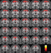
Striatal, thalamic, premotor, and prefrontal activations specific to relative, beat-based timing. BOLD activations for relative, beat-based timing (regular vs irregular) are shown in a series of coronal sections. Significant activations were found in the striatum, thalamus, premotor cortex, SMA, and prefrontal areas including the DLPFC at a threshold of p < 0.001 (uncorrected). The sections show activations at every 1 mm from y = −8 mm to = +20 mm as overlaid on the average normalized structural scan of all subjects. The strength of activations (t-value) is graded according to the color scheme at the bottom right.
Figure 5.
Figure 5.
Dissociation between neural substrates underlying absolute and relative timing. A, Glass brain image in MNI space showing activations for absolute, duration-based timing (irregular vs regular) at a t-value threshold of 4.00 and an extent threshold of 10 voxels. B, Glass brain image in MNI space showing activations for relative, beat-based timing (regular vs irregular) at a t-value threshold of 4.00 and an extent threshold of 10 voxels. C, Activations for absolute timing are depicted in yellow, and activations for relative timing are shown in green on a sagittal section of the average normalized structural image at x = +7 mm and a threshold of p < 0.01 (uncorrected) to show the clear differences in the brain bases for absolute and relative timing, respectively.

References

    1. Akkal D, Dum RP, Strick PL. Supplementary motor area and presupplementary motor area: targets of basal ganglia and cerebellar output. J Neurosci. 2007;27:10659–10673. - PMC - PubMed
    1. Artieda J, Pastor MA, Lacruz F, Obeso JA. Temporal discrimination is abnormal in Parkinson's disease. Brain. 1992;115:199–210. - PubMed
    1. Ashburner J. A fast diffeomorphic image registration algorithm. Neuroimage. 2007;38:95–113. - PubMed
    1. Barnes R, Jones MR. Expectancy, attention, and time. Cogn Psychol. 2000;41:254–311. - PubMed
    1. Belin P, Zatorre RJ, Hoge R, Evans AC, Pike B. Event-related fMRI of the auditory cortex. Neuroimage. 1999;10:417–429. - PubMed

Publication types

LinkOut - more resources