. 2011 Mar 9;31(10):3914-25.
doi: 10.1523/JNEUROSCI.3631-10.2011.

The survival of motor neuron (SMN) protein interacts with the mRNA-binding protein HuD and regulates localization of poly(A) mRNA in primary motor neuron axons

Affiliations

The survival of motor neuron (SMN) protein interacts with the mRNA-binding protein HuD and regulates localization of poly(A) mRNA in primary motor neuron axons

Claudia Fallini et al. J Neurosci. .

Abstract

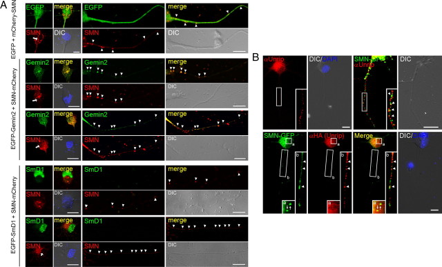
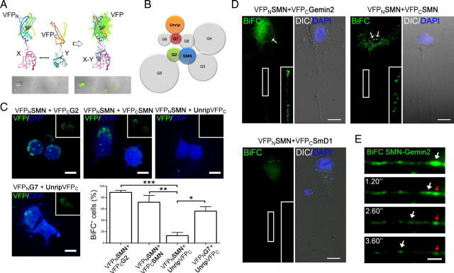
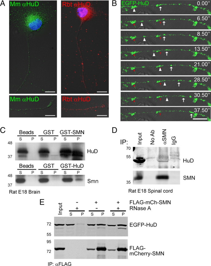
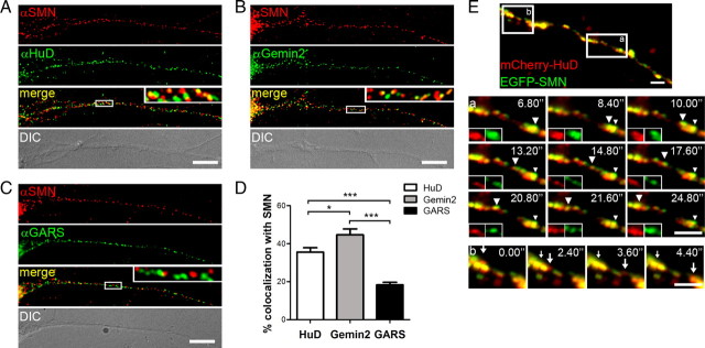
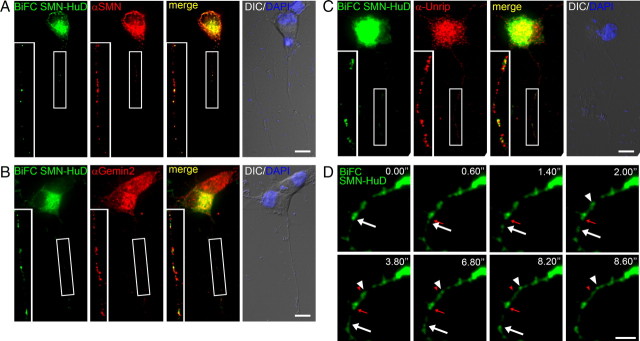
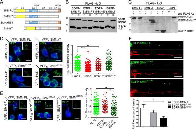
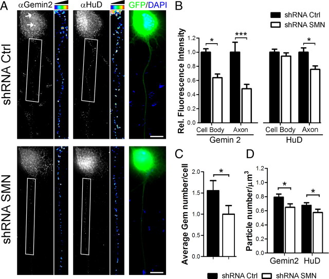
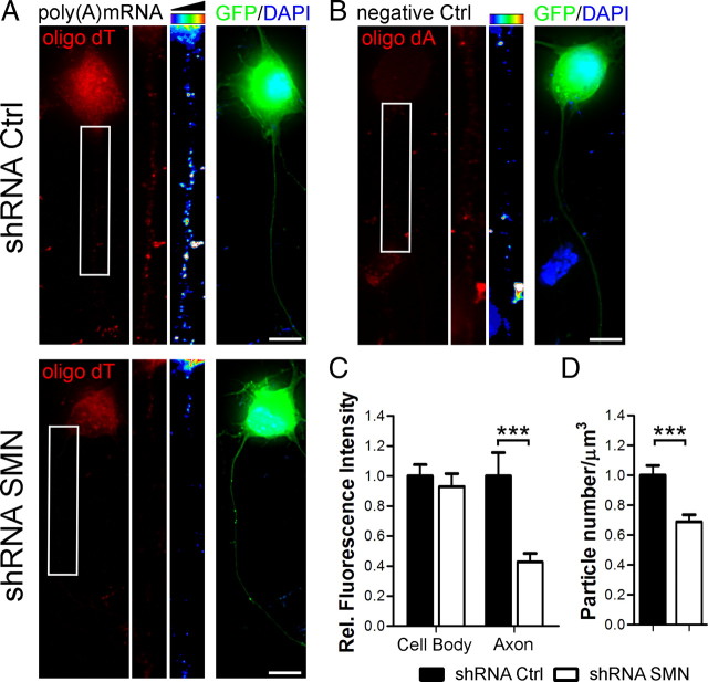
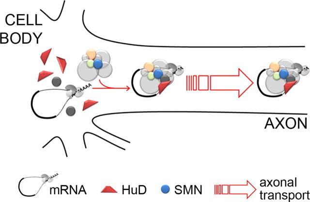
Spinal muscular atrophy (SMA) results from reduced levels of the survival of motor neuron (SMN) protein, which has a well characterized function in spliceosomal small nuclear ribonucleoprotein assembly. Currently, it is not understood how deficiency of a housekeeping protein leads to the selective degeneration of spinal cord motor neurons. Numerous studies have shown that SMN is present in neuronal processes and has many interaction partners, including mRNA-binding proteins, suggesting a potential noncanonical role in axonal mRNA metabolism. In this study, we have established a novel technological approach using bimolecular fluorescence complementation (BiFC) and quantitative image analysis to characterize SMN-protein interactions in primary motor neurons. Consistent with biochemical studies on the SMN complex, BiFC analysis revealed that SMN dimerizes and interacts with Gemin2 in nuclear gems and axonal granules. In addition, using pull down assays, immunofluorescence, cell transfection, and BiFC, we characterized a novel interaction between SMN and the neuronal mRNA-binding protein HuD, which was dependent on the Tudor domain of SMN. A missense mutation in the SMN Tudor domain, which is known to cause SMA, impaired the interaction with HuD, but did not affect SMN axonal localization or self-association. Furthermore, time-lapse microscopy revealed SMN cotransport with HuD in live motor neurons. Importantly, SMN knockdown in primary motor neurons resulted in a specific reduction of both HuD protein and poly(A) mRNA levels in the axonal compartment. These findings reveal a noncanonical role for SMN whereby its interaction with mRNA-binding proteins may facilitate the localization of associated poly(A) mRNAs into axons.


Figures

Figure 1.
Figure 1.
SMN colocalizes with Gemin2 and Unrip but not SmD1 in motor neuron axons. A, Cotransfection of an SMN-mCherry fusion construct with EGFP (top), EGFP-Gemin2 (middle), and EGFP-SmD1 (bottom) in primary motor neurons. SMN localizes to nuclear gems (left two panels, arrows), and granules in the cytoplasm and axon (right two panels, arrowheads). Axonal SMN-positive granules (arrowheads) contain Gemin2 but not SmD1, whose localization is restricted to the nuclear compartment, with few cytoplasmic granules colocalizing with SMN. B, Endogenous (top) and HA-tagged (bottom) Unrip is present in motor neuron axons and partially colocalizes with SMN-GFP in axonal granules (arrowheads) but not in nuclear gems (arrows). Insets are enlarged images of boxed sections. Scale bars, 10 μm.
Figure 2.
Figure 2.
BiFC analysis of the SMN complex. A, Schematic representation of BiFC. Two halves of VFP are tethered together by the interaction of their fusion partners, allowing the fragments to complement and reconstitute the functional fluorophore. B, BiFC was used to study SMN interactions within the classical SMN complex, as described by Otter et al. (2007). C, Neuro2a cells were cotransfected for 12 h with the BiFC pairs SMN-Gemin2, SMN-SMN, SMN-Unrip, and Gemin7-Unrip, and VFP mean fluorescence intensity was measured for each individual cell. Values >1.5-fold the background were considered positive (BiFC+; one-way ANOVA and Tukey's post hoc test, n = 168, 150, 155, 198 for SMN-G2, SMN-SMN, SMN-Unrip, and Unrip-G7, respectively; *p < 0.05, **p < 0.01, ***p < 0.001). CFP was used to identify transfected cells for the analysis. Insets show the VFP signal alone. D, BiFC was used in primary motor neurons to visualize the cellular localization of interactions. SMN-Gemin2 and SMN-SMN pairs show BiFC signal in both nuclear gems (arrows) and axonal granules (insets). SMN-SmD1 BiFC-positive granules are restricted to cell body and nucleus. E, Live cell imaging of an SMN-Gemin2 BiFC granule in the axon of primary cultured motor neuron. The granule (arrows) moved for 8.66 μm at an average speed of 3.09 μm/s (starting position is indicated by the red arrows). The apparent reduction in the size of the granules over time is due to VFP photobleaching during the movie acquisition. Scale bars: C, E, 5 μm; D, 10 μm.
Figure 3.
Figure 3.
The mRNA-binding protein HuD localizes to motor neuron axons and is copurified with SMN. A, Endogenous HuD was detected by immunostaining using a monoclonal mouse (Mm, left) or a polyclonal rabbit antibody (Rbt, right). HuD-positive granules were visible in the cell body and proximal axon (top), as well as in the growth cones (bottom). DAPI was used to stain nuclei (blue). B, EGFP-HuD was expressed in primary motor neurons and active transport of granules was imaged by time lapse microscopy. The marked granules moved anterogradely toward the growth cone at an average speed of 0.75 μm/s (arrowheads) and 0.98 μm/s (arrows). Images of the fluorescent signals have been overlaid on DIC images in both A and B. Scale bars: A, 10 μm; B, 5 μm. C, Endogenous HuD (top) and SMN (bottom) were copurified from rat brain extracts using GST-fusions of SMN (top) and HuD (bottom). GST (lanes 3 and 4) or beads alone (lanes 5 and 6) were used as negative controls. D, Endogenous SMN protein was immunoprecipitated from E18 rat spinal cord lysates and the pellet was immunoblotted for HuD (top, lane 3) and SMN (bottom). Mouse IgG (lane 4) or no antibody (lane 2) were used as controls. Immunoprecipitation input is shown in lane 1. E, EGFP-HuD and FLAG-mCherry-SMN expressed in HEK293 cells were coprecipitated using an anti-FLAG antibody. A band corresponding to EGFP-HuD was enriched in the immunoprecipitation pellet (P) and present in the supernatant (S) when FLAG-mCherry-SMN was coexpressed (lanes 4–5). Pretreatment with 1 mg/ml RNase A for 1 h did not affect the interaction (lanes 7–8). Cells expressing EGFP-HuD alone (lanes 2–3) were used as a negative control. Immunoprecipitation input is shown (lane 1).
Figure 4.
Figure 4.
SMN and HuD colocalize and are cotransported in the same granule in motor neurons. A–C, Endogenous SMN and HuD (A), Gemin2 (B), and GARS (C) proteins were detected by immunofluorescence in primary motor neurons. Strong colocalization was observed between SMN and HuD and SMN and Gemin2, but not between SMN and GARS (insets). D, Quantification of colocalization between each protein and SMN was performed. Bars represent mean and SEM (one-way ANOVA and Tukey's post hoc test, n = 30; *p < 0.05, ***p < 0.001). E, Dual fluorescent-tagged SMN and HuD are cotransported in the same granule in the axon of motor neuron. The SMN-HuD colocalized granule shown in Ea (arrowheads) moved for 13.74 μm with an average speed of 0.88 μm/s. Single-channel magnifications of the granule are shown in the insets. A granule positive for mCherry-HuD alone is shown in Eb (arrows). Small arrowheads and arrows mark the initial position of the tracked granules. Scale bars: A–C, 10 μm; E, 5 μm.
Figure 5.
Figure 5.
BiFC reveals SMN-HuD granules localized and actively transported along motor neuron axons. A–C, Primary motor neurons were transfected with SMN and HuD BiFC constructs and fixed 24 h posttransfection. BiFC granules (green) were stained using anti-SMN (A), Gemin2 (B), and Unrip (C) antibodies (red). Positive granules were visible in the cell body and along the axon (insets). DAPI (blue) was used to identify nuclei. D, Time lapse microscopy was used to image SMN-HuD BiFC granules moving in motor neuron processes. Anterograde (arrows) and retrograde (arrowheads) trajectories were observed. Both granules moved at an average speed of 1 μm/s. Red arrowheads and arrows mark the initial position of the tracked granules. Scale bars: A–C, 10 μm; D, 5 μm.
Figure 6.
Figure 6.
Deletion and mutation in the SMN Tudor domain specifically impair SMN-HuD interaction. A, Schematic representation of SMN deletion constructs lacking exon 7 (SMNΔ7), the N terminus (SMNΔN53), or the Tudor domain (SMNΔT). The E134K (SMNE134K) or the G279V (SMNG279V) mutations were introduced in the murine full-length SMN (SMN FL) sequence. B, C, Immunoprecipitation (IP) experiments with HEK293 cells transfected with EGFP- and FLAG-tagged SMN and HuD constructs. Anti-FLAG antibody was used to precipitate FLAG-tagged HuD. Monoclonal antibodies to GFP and FLAG were used for detection. SMN with the Tudor domain deletion (SMNΔT) failed to coprecipitate with HuD (B, lane 7), in contrast to SMN full-length and N- or C-terminal deletions. The Tudor domain alone, fused to EGFP, is sufficient to be copurified with FLAG-HuD (C, lane 5). P, IP pellet; S, IP supernatant. D, E, BiFC was used to investigate the effect of Tudor domain deletion or mutation on SMN-HuD interaction (D) and SMN self-association (E). Neuro2a cells were transfected with BiFC constructs and fixed after 12 h. Murine full-length SMN (SMN FL), SMNΔT, and Tudor domain (SMNE134K) or YG-box mutants (SMNG279V) were compared. Fluorescence intensity of the BiFC signal (green) was quantified and normalized to CFP protein expression (blue). Both deletion (SMNΔT) and mutation (SMNE134K) in the Tudor domain, but not in the YG-box (SMNG279V), showed significantly reduced interactions with HuD (D, graph). In contrast, SMNG279V mutant showed impaired ability to self-associate compared with both SMN FL and the SMNE134K constructs (E, graph). Scatter plots represent the quantification of 80–120 cells per condition from three independent experiments. Mean and SEM are shown (one-way ANOVA and Tukey's post hoc test, **p < 0.01, ***p < 0.001). F, Primary motor neurons were transfected with EGFP-fusions of wild-type or mutant SMN cDNAs (SMN FL, SMNE134K, and SMNG279V respectively; green), and stained with tau antibody (red) to recognize the axons. EGFP fluorescence intensity was evaluated in the proximal axonal segment. A significant reduction was observed when the G279V mutation was present (one-way ANOVA and Tukey's post hoc test, n = 21, 24, and 27 for SMN FL, SMNE134K, and SMNG279V respectively; *p < 0.05). Scale bars: D–F, 10 μm.
Figure 7.
Figure 7.
SMN deficiency impairs HuD axonal localization. A, Primary motor neurons were transfected with an shRNA construct targeting SMN mRNA (bottom) or a nonsilencing control (top). Cells were fixed after 5 d and stained for Gemin2 (left) and HuD (right) proteins. GFP expression (green) was used to identify transfected motor neurons. Nuclei were stained with DAPI (blue). Axons were straightened and pseudo-colored with an intensity map (insets). Gems are indicated by the arrows. Scale bar, 10 μm. B, D, Gemin2 and HuD average fluorescence intensity (B) and particle number (D) were quantified in the proximal axonal fragment (B, D) and cell body (B, only) of shRNA SMN and control (Ctrl) cells from three independent experiments. While Gemin2 protein was significantly decreased in both cell compartments, HuD levels were selectively reduced in the axons by 24% compared with controls (one-way ANOVA and Tukey's post hoc test, n = 28, *p < 0.5, ***p < 0.001). C, Gems, identified as Gemin2-positive bodies in the nucleus, were scored. Gem number decreased from 1.5 ± 0.3 in control cells to 1 ± 0.7 gem/cell in knockdown motor neurons (Student's t test, n = 28, *p < 0.05). For all graphs, columns represent mean and SEM.
Figure 8.
Figure 8.
SMN knockdown impairs the localization of poly(A)-positive mRNA granules in motor neuron axons. A, B, Primary motor neurons were transfected with an shRNA construct targeting SMN mRNA (bottom) or a nonsilencing control (top). Cells were fixed after 5 d and processed for fluorescence in situ hybridization. Poly(A)+ mRNAs were detected using a DIG-labeled oligo dT probe (red) (A). The specificity of the signal was controlled by using an oligo dA probe (B). GFP expression (green) was used to identify transfected motor neurons. Nuclei were stained with DAPI (blue). Axons were straightened and pseudo-colored with a 16-color intensity map (insets). Scale bar, 10 μm. C, D, Average fluorescence intensity (C) and number (D) of poly(A)+ mRNA-containing granules were quantified in the proximal axonal fragment and cell body of shRNA SMN and control (Ctrl) cells from three independent experiments. Both parameters were significantly reduced in respect to controls by 58% and 31%, respectively (one-way ANOVA and Tukey's post hoc test, n = 31 for shRNA Ctrl and n = 36 shRNA SMN respectively, ***p < 0.001). Bars represent mean and SEM.
Figure 9.
Figure 9.
Proposed model for SMN function in motor neuron axons. SMN, either alone or associated with components of the SMN complex, facilitates the association of HuD and possibly other mRNA-binding proteins with target mRNAs and the trafficking of associated poly(A) mRNAs from the cell body along the motor neuron axon.

References

    1. Akamatsu W, Fujihara H, Mitsuhashi T, Yano M, Shibata S, Hayakawa Y, Okano HJ, Sakakibara S, Takano H, Takano T, Takahashi T, Noda T, Okano H. The RNA-binding protein HuD regulates neuronal cell identity and maturation. Proc Natl Acad Sci U S A. 2005;102:4625–4630. - PMC - PubMed
    1. Anthony K, Gallo JM. Aberrant RNA processing events in neurological disorders. Brain Res. 2010;1338:67–77. - PubMed
    1. Aronov S, Aranda G, Behar L, Ginzburg I. Axonal tau mRNA localization coincides with tau protein in living neuronal cells and depends on axonal targeting signal. J Neurosci. 2001;21:6577–6587. - PMC - PubMed
    1. Aronov S, Aranda G, Behar L, Ginzburg I. Visualization of translated tau protein in the axons of neuronal P19 cells and characterization of tau RNP granules. J Cell Sci. 2002;115:3817–3827. - PubMed
    1. Atlas R, Behar L, Elliott E, Ginzburg I. The insulin-like growth factor mRNA binding-protein IMP-1 and the Ras-regulatory protein G3BP associate with tau mRNA and HuD protein in differentiated P19 neuronal cells. J Neurochem. 2004;89:613–626. - PubMed

Publication types

MeSH terms