. 2011 Jun;25(6):2001-11.
doi: 10.1096/fj.10-177378. Epub 2011 Mar 9.

Cardiopulmonary function in two human disorders of the hypoxia-inducible factor (HIF) pathway: von Hippel-Lindau disease and HIF-2alpha gain-of-function mutation

Affiliations

Cardiopulmonary function in two human disorders of the hypoxia-inducible factor (HIF) pathway: von Hippel-Lindau disease and HIF-2alpha gain-of-function mutation

Federico Formenti et al. FASEB J. 2011 Jun.

Abstract

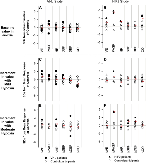
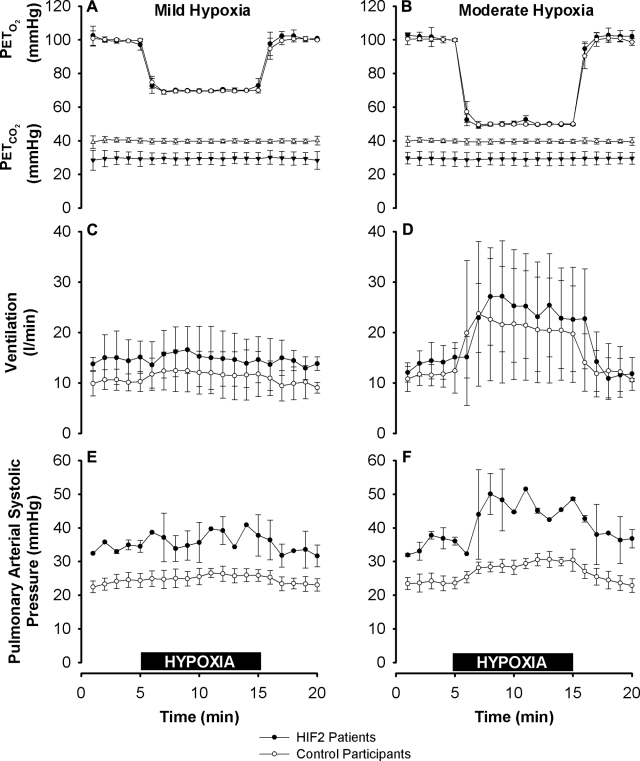
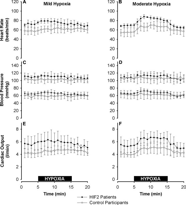
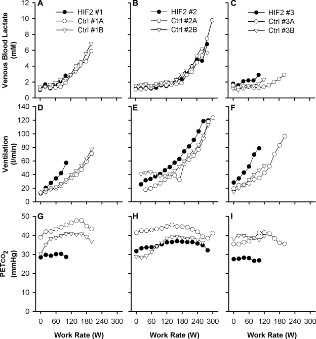
The hypoxia-inducible factors (HIFs; isoforms HIF-1α, HIF-2α, HIF-3α) mediate many responses to hypoxia. Their regulation is principally by oxygen-dependent degradation, which is initiated by hydroxylation of specific proline residues followed by binding of von Hippel-Lindau (VHL) protein. Chuvash polycythemia is a disorder with elevated HIF. It arises through germline homozygosity for hypomorphic VHL alleles and has a phenotype of hematological, cardiopulmonary, and metabolic abnormalities. This study explores the phenotype of two other HIF pathway diseases: classic VHL disease and HIF-2α gain-of-function mutation. No cardiopulmonary abnormalities were detected in classic VHL disease. HIF-2α gain-of-function mutations were associated with pulmonary hypertension, increased cardiac output, increased heart rate, and increased pulmonary ventilation relative to metabolism. Comparison of the HIF-2α gain-of-function responses with data from studies of Chuvash polycythemia suggested that other aspects of the Chuvash phenotype were diminished or absent. In classic VHL disease, patients are germline heterozygous for mutations in VHL, and the present results suggest that a single wild-type allele for VHL is sufficient to maintain normal cardiopulmonary function. The HIF-2α gain-of-function phenotype may be more limited than the Chuvash phenotype either because HIF-1α is not elevated in the former condition, or because other HIF-independent functions of VHL are perturbed in Chuvash polycythemia.


Figures

Figure 1.
Figure 1.
Cardiopulmonary variables in both the VHL and HIF2 studies: baseline measurements (A, B) and incremental responses with mild hypoxia (C, D) and moderate hypoxia (E, F). Results are shown in terms of number of (control group) sd by which each participant's baseline and response differed from the mean baseline and mean response, respectively, of the control participants. Red symbols show average results for patient groups. A, C, E) VHL patients were not significantly different from the control participants for any variable. B, D, F) Under baseline conditions, HIF2 patients had significantly higher pulmonary arterial pressure, heart rate, and cardiac output than control participants. Compared with control participants, HIF2 patients also had a significantly greater rise in pulmonary arterial pressure with moderate hypoxia. VE, ventilation; PASP, pulmonary arterial systolic pressure; HR, heart rate; SBP, systolic blood pressure; DBP, diastolic blood pressure; CO, cardiac output; Δ, increment.
Figure 2.
Figure 2.
End-tidal gas control, ventilation, and pulmonary arterial systolic pressure during mild and moderate hypoxia. A, B) PETo2 and PETco2 in mild hypoxia (A) and moderate hypoxia (B). PETo2 was well matched between groups. PETco2 was lower for the HIF2 patients, reflecting their lower baseline air-breathing PETco2. C, D) Ventilation, given at body temperature and pressure, saturated with water vapor, in mild (C) and moderate hypoxia (D). Baseline ventilation and the increase in ventilation provoked by hypoxia were not significantly different between the two groups. E, F) Pulmonary arterial systolic pressure. Compared with control participants, HIF2 patients had elevated pulmonary arterial systolic pressures at baseline (24±2 vs. 35±1 mmHg, P<0.003), and increased sensitivity to moderate (F) but not mild (E) hypoxia (5.5±1.3 vs. 11.5±0.7 mmHg, P<0.004). Values are expressed as means ± sd. Horizontal black bars indicate 10-min periods of mild (A, C, E) and moderate (B, D, F) exposures to hypoxia.
Figure 3.
Figure 3.
Systemic vascular responses in HIF2 patients and control participants to mild and moderate hypoxia. A, B) Heart rate in mild hypoxia (A) and moderate hypoxia (B). At baseline, heart rate was significantly higher in the HIF2 patients than in the control participants (P<0.02). C, D) Systolic (top) and diastolic (bottom) blood pressure in mild (C) and moderate hypoxia (D). Blood pressure did not differ significantly between the two groups. E, F) Cardiac output, assessed noninvasively using Doppler echocardiography, in mild (E) and moderate hypoxia (F). At baseline, cardiac output was significantly greater in the HIF2 patients than in the control participants (P<0.02). Increments in heart rate, blood pressure, and cardiac output with hypoxia did not differ significantly between the two groups. Values are expressed as means ± sd. Horizontal black bars indicate 10-min periods of mild (A, C, E) and moderate (B, D, F) exposures to hypoxia.
Figure 4.
Figure 4.
Individual responses for HIF2 patients and control participants during the incremental exercise test to exhaustion. A–C) Venous blood lactate concentration as a function of work rate. D–F) Ventilation as a function of work rate. G–I) PETco2 as a function of work rate. Results are for HIF2 patient 1 (A, D, G), HIF2 patient 2 (B, E, H), and HIF2 patient 3 (C, F, I) and their associated control participants. Data are minute averages.

References

    1. Schofield C. J., Ratcliffe P. J. (2004) Oxygen sensing by HIF hydroxylases. Nat. Rev. Mol. Cell. Biol. 5, 343–354 - PubMed
    1. Semenza G. L. (2004) Hydroxylation of HIF-1: oxygen sensing at the molecular level. Physiology 19, 176–182 - PubMed
    1. Semenza G. L. (2007) Oxygen-dependent regulation of mitochondrial respiration by hypoxia-inducible factor 1. Biochem. J. 405, 1–9 - PubMed
    1. Wenger R. H. (2002) Cellular adaptation to hypoxia: O2-sensing protein hydroxylases, hypoxia-inducible transcription factors, and O2-regulated gene expression. FASEB J. 16, 1151–1162 - PubMed
    1. Wang G. L., Jiang B. H., Rue E. A., Semenza G. L. (1995) Hypoxia-inducible factor 1 is a basic-helix-loop-helix-PAS heterodimer regulated by cellular O2 tension. Proc. Natl. Acad. Sci. U. S. A. 92, 5510–5514 - PMC - PubMed

Publication types

MeSH terms

Substances