. 2011 Mar 31;471(7340):656-60.
doi: 10.1038/nature09795. Epub 2011 Mar 9.

The structural basis of agonist-induced activation in constitutively active rhodopsin

Affiliations

The structural basis of agonist-induced activation in constitutively active rhodopsin

Jörg Standfuss et al. Nature. .

Abstract

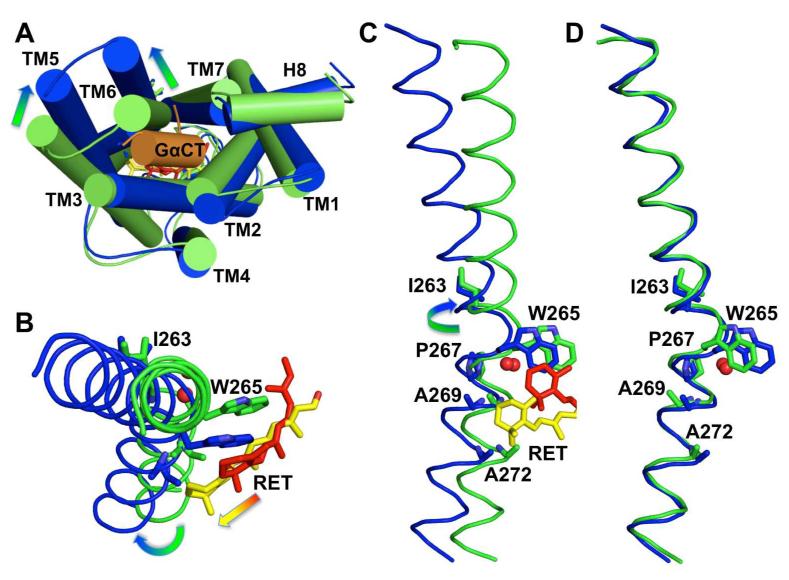
G-protein-coupled receptors (GPCRs) comprise the largest family of membrane proteins in the human genome and mediate cellular responses to an extensive array of hormones, neurotransmitters and sensory stimuli. Although some crystal structures have been determined for GPCRs, most are for modified forms, showing little basal activity, and are bound to inverse agonists or antagonists. Consequently, these structures correspond to receptors in their inactive states. The visual pigment rhodopsin is the only GPCR for which structures exist that are thought to be in the active state. However, these structures are for the apoprotein, or opsin, form that does not contain the agonist all-trans retinal. Here we present a crystal structure at a resolution of 3 Å for the constitutively active rhodopsin mutant Glu 113 Gln in complex with a peptide derived from the carboxy terminus of the α-subunit of the G protein transducin. The protein is in an active conformation that retains retinal in the binding pocket after photoactivation. Comparison with the structure of ground-state rhodopsin suggests how translocation of the retinal β-ionone ring leads to a rotation of transmembrane helix 6, which is the critical conformational change on activation. A key feature of this conformational change is a reorganization of water-mediated hydrogen-bond networks between the retinal-binding pocket and three of the most conserved GPCR sequence motifs. We thus show how an agonist ligand can activate its GPCR.


Figures

Figure 1
Figure 1. Conformational changes in the retinal binding pocket
A: 2Fo-Fc map (contoured at 1.5 sigma) of the retinal-binding pocket. The retinal β-ionone ring is well resolved while density of the polyene chain broadens towards the end facing K2967.43. Occupancy refinements indicated a mixture of 60% all-trans-retinal and 40% of isomers rotated around single bonds or double bonds after position C9. An acetate molecule is packed between F2085.43 and F2766.59 and blocks a potential retinal entry/exit channel. B: Superposition of E113Q/GαCT (blue, 2×72) with ground state rhodopsin (green, 1GZM). Compared to the β-ionone ring of 11-cis-retinal (red) in ground state rhodopsin, the β-ionone ring of all-trans-retinal (yellow) in the active E113Q/GαCT structure is shifted 4.3 Å towards the cleft between TM5 and TM6, where it makes contact with both helices. Simultaneously W2656.48 of the CWxP motif is released from its locked position in the ground state, which disrupts a water-mediated interaction to S2987.45 and breaks the restraining TM6-TM7 link. The salt bridge between E1133.28 as counterion to the protonated Schiff base and K2967.43 is broken in the E113Q/GαCT structure removing a link known to restrain TM3 and TM7 in the inactive, ground-state conformation. By alteration of these three key interactions retinal can induce the rotation of TM6 that opens the G protein-binding site and simultaneously can facilitate a reorganization of hydrogen-bonding networks in the NPxxY and E(D)RY at the cytoplasmic end of TM3 and TM7.
Figure 2
Figure 2. Rearrangement of the heptahelix bundle and rotation of TM6
A: Superposition of Cα traces of ground state rhodopsin (green) and E113Q (blue) with bound G protein peptide (orange), as seen from the cytoplasmic side. The main rearrangements of the seven transmembrane helices (cylinders) that open the G protein-binding site are indicated as arrows. The loop regions have been smoothed for clarity. B, C: Cytoplasmic and membrane side views of TM6 (Cα traces in ribbon representation) from ground state rhodopsin and the E113Q/GαCT structure illustrates how 11-cis (red) to all-trans (yellow) isomerisation of retinal can release W2656.48 from its locked position in the ground state and insert the β-ionone ring between F2085.43/F2125.47 in H5 and A2696.52/A2726.55 in H6. This leads to a rotation of TM6 that is amplified towards its cytoplasmic end by the characteristic bend of the helix introduced by P2676.50 and water7. D: Superposition of TM6 alone shows that the shape of the helix is preserved during the rearrangements.
Figure 3
Figure 3. Rearrangement of water mediated hydrogen-bonding networks
A: The E113Q structure reveals potential water mediated hydrogen-bonding networks that connect the retinal-binding region with the GαCT binding site. Strong electron density (Blue mesh represents 2Fo-Fc map contoured at 2.0 sigma. Green mesh shows the Fo-Fc map contoured at 3.0 sigma) difference peaks are observed when waters are omitted during simulated annealing refinement, demonstrating a high degree of local order. B: In ground-state rhodopsin, the hydrogen-bonding network connects W2656.48 in the retinal-binding pocket with N3027.49 of the NPxxY motif (blue) and the carboxyl of M2576.40, via the conserved residues S2987.45 and D832.50. M2576.40 is part of a region called the hydrophobic barrier (green) that separates the ionic lock interaction between the E1343.49/R1353.50 pair in the E(D)RY motif (salmon) of TM3 and E2476.30 in TM6. C: In the active E113Q/GαCT structure, TM6 is rotated including W2656.48, which breaks the water-mediated link to S2987.45 in TM7. The TM1-TM2-TM7 network between N551.50, D832.50 and S2987.45 is reorganized but maintains and stabilizes a similar distortion of TM7 induced by P3037.50 of the NPxxY motif (blue). The hydrophobic barrier that separates the E(D)RY motif (salmon) in TM3 from the membrane core is opened. Y3067.53 of the NPxxY motif (blue) in TM7 and Y2235.58 in TM5 rearrange to fill the resulting gap and extend the hydrogen-bonded network towards R1353.50 of the E(D)RY motif (salmon). R1353.50 is released from its ground state bent interaction with E1343.49, which opens the ionic lock and facilitates the outward movement of TM6 that allows binding of the GαCT peptide (orange ribbon). In the indicated location the water cluster can bridge the retinal-binding core with the cytoplasmic side of the receptor and is directly mediating interactions to the G protein peptide.
Figure 4
Figure 4. Activation of rhodopsin by the agonist all-trans-retinal
The protein backbone of E113Q/GαCT is shown in cyan with predominant conformational changes in TM5 and TM6 (RMSD of Cα atoms with respect to 1GZM > 3.5Å) in blue. The key regions involved in rhodopsin activation and discussed in the text are highlighted. The side-chains of the CWxP motif close to the retinal-binding site are coloured red. Side-chains of the NPxxY motif are coloured, blue and extend the hydrogen-bonding network through the green hydrophobic barrier. Side-chains of the E(D)RY motif, as part of the ionic lock and the G protein-binding site are coloured salmon. The GαCT peptide is shown as an orange ribbon. The engineered disulfide bond in the extracellular domain is well isolated from the structural motifs involved in rhodopsin activation, an explanation for its neutral stabilizing characteristics.

References

    1. Rosenbaum DM, Rasmussen SG, Kobilka BK. The structure and function of G-protein-coupled receptors. Nature. 2009;459(7245):356. - PMC - PubMed
    1. Park JH, et al. Crystal structure of the ligand-free G-protein-coupled receptor opsin. Nature. 2008;454(7201):183. - PubMed
    1. Scheerer P, et al. Crystal structure of opsin in its G-protein-interacting conformation. Nature. 2008;455(7212):497. - PubMed
    1. Zhukovsky EA, Oprian DD. Effect of carboxylic acid side chains on the absorption maximum of visual pigments. Science. 1989;246(4932):928. - PubMed
    1. Sakmar TP, Franke RR, Khorana HG. Glutamic acid-113 serves as the retinylidene Schiff base counterion in bovine rhodopsin. Proc Natl Acad Sci U S A. 1989;86(21):8309. - PMC - PubMed

Publication types

MeSH terms

Associated data