. 2011 Mar 10;471(7337):240-4.
doi: 10.1038/nature09773.

DNA ligase III is critical for mtDNA integrity but not Xrcc1-mediated nuclear DNA repair

Affiliations

DNA ligase III is critical for mtDNA integrity but not Xrcc1-mediated nuclear DNA repair

Yankun Gao et al. Nature. .

Abstract

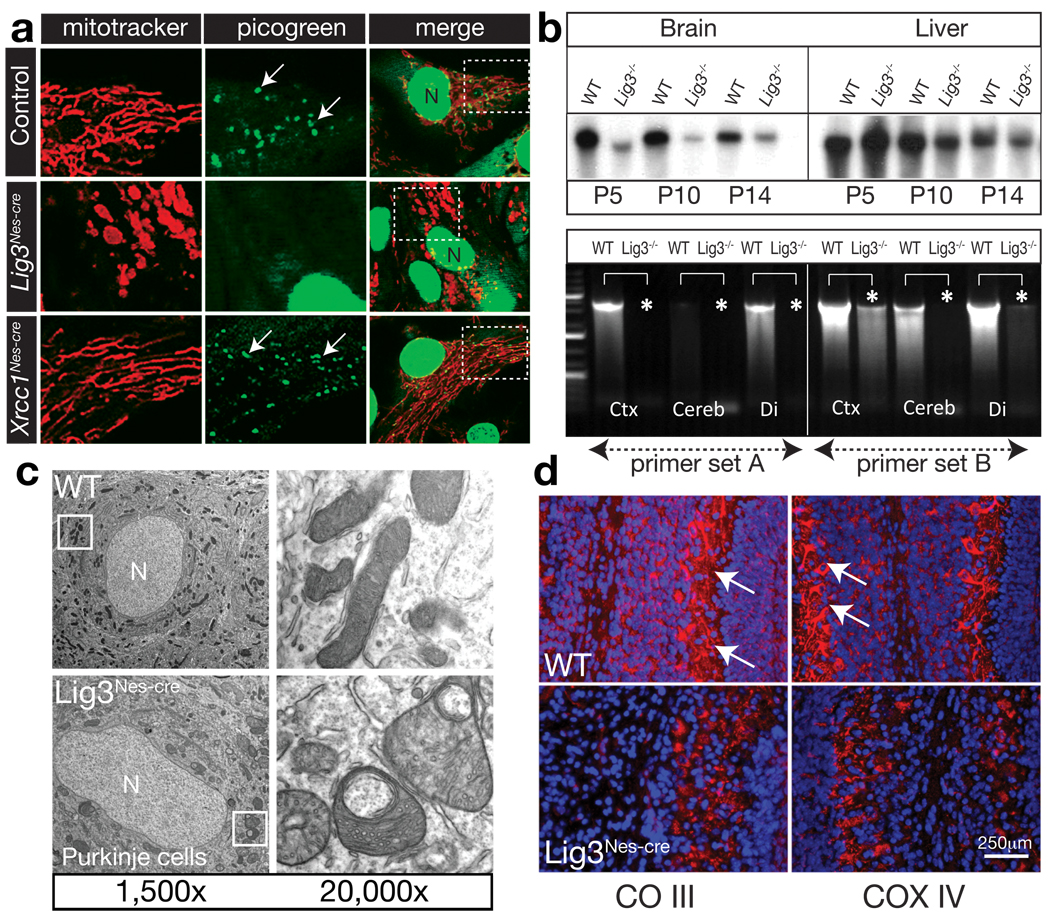
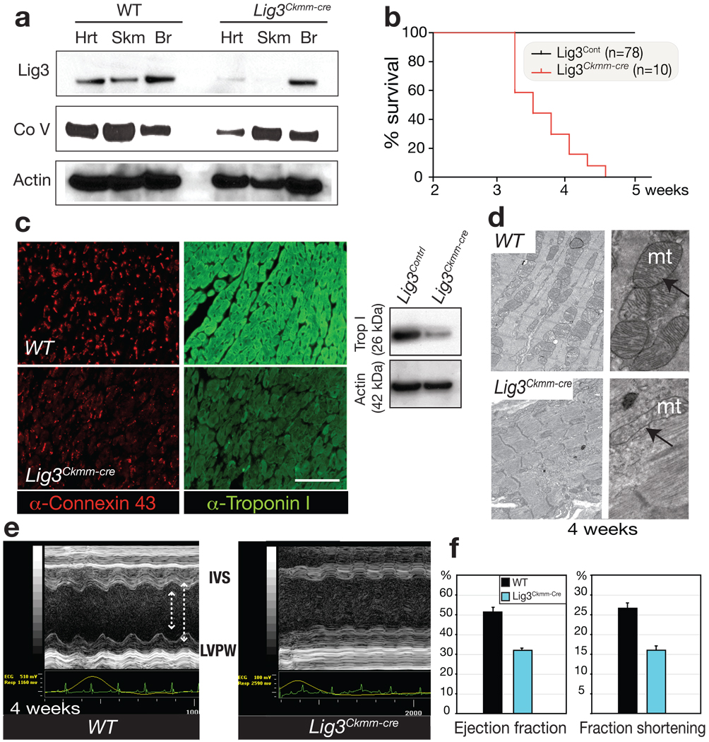
DNA replication and repair in mammalian cells involves three distinct DNA ligases: ligase I (Lig1), ligase III (Lig3) and ligase IV (Lig4). Lig3 is considered a key ligase during base excision repair because its stability depends upon its nuclear binding partner Xrcc1, a critical factor for this DNA repair pathway. Lig3 is also present in the mitochondria, where its role in mitochondrial DNA (mtDNA) maintenance is independent of Xrcc1 (ref. 4). However, the biological role of Lig3 is unclear as inactivation of murine Lig3 results in early embryonic lethality. Here we report that Lig3 is essential for mtDNA integrity but dispensable for nuclear DNA repair. Inactivation of Lig3 in the mouse nervous system resulted in mtDNA loss leading to profound mitochondrial dysfunction, disruption of cellular homeostasis and incapacitating ataxia. Similarly, inactivation of Lig3 in cardiac muscle resulted in mitochondrial dysfunction and defective heart-pump function leading to heart failure. However, Lig3 inactivation did not result in nuclear DNA repair deficiency, indicating essential DNA repair functions of Xrcc1 can occur in the absence of Lig3. Instead, we found that Lig1 was critical for DNA repair, but acted in a cooperative manner with Lig3. Additionally, Lig3 deficiency did not recapitulate the hallmark features of neural Xrcc1 inactivation such as DNA damage-induced cerebellar interneuron loss, further underscoring functional separation of these DNA repair factors. Therefore, our data reveal that the critical biological role of Lig3 is to maintain mtDNA integrity and not Xrcc1-dependent DNA repair.


Figures

Figure 1
Figure 1. Lig3 inactivation throughout the nervous system leads to a phenotype different to Xrcc1 loss
(a) MRI analysis of the Lig3Nes-cre nervous system shows microcephaly and a small cerebellum (arrow) compared to WT. (b) Lig3 is deleted in various Lig3Nes-cre brain regions while Xrcc1 protein levels are unaffected. (c) Comparative Nissl staining of P14 cerebellum (Ce) from Lig3Nes-cre, Xrcc1Nes-cre and WT. (d) PCNA immunostaining shows disrupted neurogenesis in the P6 Lig3Nes-cre cerebellum (arrows). (e) Levels of the interneuron marker Pax2 are similar in control and Lig3Nes-cre tissue, but substantially reduced in the Xrcc1Nes-cre cerebellum. (f) Interneurons are present in the Lig3Nes-cre cerebellum but are absent from Xrcc1Nes-cre tissue (arrows) as determined by Pax2 immunostaining.
Figure 2
Figure 2. Mitochondrial function is disrupted in the Lig3Nes-cre brain
(a) Mitotracker labeling reveals defects in mitochondria from Lig3Nes-cre but not WT or Xrcc1Nes-cre astrocytes. Picogreen staining shows reduced mtDNA in Lig3Nes-cre astrocytes; N is the nucleus. Boxed areas show the region expanded in adjacent panels. (b) Analysis of mtDNA via pulse field gel electrophoresis shows an age-dependent quantitative reduction in mtDNA from P5-P14 Lig3Nes-cre brain compared with controls. mtDNA levels in the Lig3Nes-cre liver were similar to control tissue. PCR of mtDNA revealed a reduction in mtDNA isolated from Lig3Nes-cre brain tissue (asterisks). Ctx is cortex, Cereb is cerebellum and Di is diencephalon. (c) Electron microscopy showed alterations in mitochondrial morphology in Lig3-deficient Purkinje cells compared to WT tissue. (d) Electron transport chain components complex III core 2 (CO III) and COX IV staining in vivo is reduced in P7 cerebellar Purkinje cell mitochondria; counterstain is DAPI.
Figure 3
Figure 3. Lig3 inactivation causes cardiac failure associated with defective mitochondrial function
(a) Lig3 protein levels are reduced in Lig3Ckmm-cre heart and skeletal muscle. A reduction of complex V subunit α also occurs in Lig3Ckmm-cre heart. (b) Lig3Ckmm-cre mice die between 3.5 and 4.5 weeks of age; n is the number of animals. (c) Altered connexin-43 distribution and depletion of cardiac troponin I occurs in 4-week-old Lig3Ckmm-cre heart. A Western blot shows decreased cardiac troponin I levels. (d) Electron microscopy of cardiac muscle shows disruption in myofiber structure and defects in mitochondrial morphology. (e) Echocardiographic analysis of Lig3Ckmm-cre mice shows movement of the interventricular septum (IVS) and the posterior walls of the left ventricle (LVPW) were decreased in the Lig3Ckmm-Cre heart (arrows). Traces below the echocardiogram show ECG readings indicate abnormal heart rhythm. (f) Cardiac contractility was also significantly depressed with decreased ejection fraction and fraction shortening indicating that the pump function of the Lig3Ckmm-Cre heart is defective. Three individual animals of each genotype were used; error bars represent standard deviation.
Figure 4
Figure 4. Lig3 is not essential for nuclear DNA repair
(a) Alkaline comet analysis shows DNA strand break repair after hydrogen peroxide treatment is similar between WT and Lig3Nes-cre quiescent primary astrocytes, but is defective in Xrcc1Nes-cre cells, as is repair after ionizing radiation (b); R30/R60 is recovery from the genotoxin after 30 or 60 minutes. Assays were run in triplicate from at least three separate experiments; error bars in all panels represent standard error of the mean. (c) Western blot analysis indicates specific shRNA-mediated knockdown of mammalian DNA ligases. Non-target scrambled (Scr) and Nbs1 shRNAs were included as controls, and Ponceau staining indicates relative protein transfer. (d) Lig1 is required for DNA repair after H2O2 damage; dual inactivation of Lig1 and Lig3 reveals cooperation between these ligases during repair. Tdp1−/− MEFS are a repair-deficient control. Photograph indicates cells are quiescent based on p27+ and PCNA immunoreactivity. (e) Representative individual comet tail moments of shRNA-mediated knockdowns after H2O2 treatment. (f) Lig1 and Lig4 are required for DNA repair after IR. Lig1 and Lig3 cooperate in the repair of IR-induced DNA damage. (g) DNA repair competency after H2O2 treatment is restored using a shLig1-resistant version of Lig1 cDNA (Lig1shR).

References

    1. Ellenberger T, Tomkinson AE. Eukaryotic DNA ligases: structural and functional insights. Annu Rev Biochem. 2008;77:313–338. - PMC - PubMed
    1. Caldecott KW, McKeown CK, Tucker JD, Ljungquist S, Thompson LH. An interaction between the mammalian DNA repair protein XRCC1 and DNA ligase III. Mol Cell Biol. 1994;14:68–76. - PMC - PubMed
    1. Ljungquist S, Kenne K, Olsson L, Sandstrom M. Altered DNA ligase III activity in the CHO EM9 mutant. Mutat Res. 1994;314:177–186. - PubMed
    1. Lakshmipathy U, Campbell C. Mitochondrial DNA ligase III function is independent of Xrcc1. Nucleic Acids Res. 2000;28:3880–3886. - PMC - PubMed
    1. Puebla-Osorio N, Lacey DB, Alt FW, Zhu C. Early embryonic lethality due to targeted inactivation of DNA ligase III. Mol Cell Biol. 2006;26:3935–3941. - PMC - PubMed

Publication types

MeSH terms