. 2011 Mar;13(3):206-16.
doi: 10.1593/neo.101332.

Vascular CD39/ENTPD1 directly promotes tumor cell growth by scavenging extracellular adenosine triphosphate

Affiliations

Vascular CD39/ENTPD1 directly promotes tumor cell growth by scavenging extracellular adenosine triphosphate

Lili Feng et al. Neoplasia. 2011 Mar.

Abstract

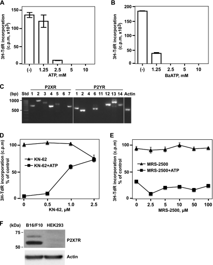
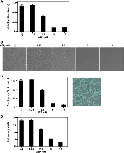
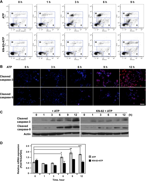
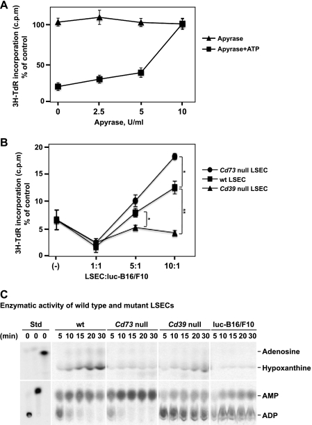
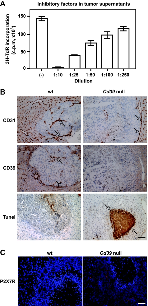
Extracellular adenosine triphosphate (ATP) is known to boost immune responses in the tumor microenvironment but might also contribute directly to cancer cell death. CD39/ENTPD1 is the dominant ectonucleotidase expressed by endothelial cells and regulatory T cells and catalyzes the sequential hydrolysis of ATP to AMP that is further degraded to adenosine by CD73/ecto-5'-nucleotidase. We have previously shown that deletion of Cd39 results in decreased growth of transplanted tumors in mice, as a result of both defective angiogenesis and heightened innate immune responses (secondary to loss of adenosinergic immune suppression). Whether alterations in local extracellular ATP and adenosine levels as a result of CD39 bioactivity directly affect tumor growth and cytotoxicity has not been investigated to date. We show here that extracellular ATP exerts antitumor activity by directly inhibiting cell proliferation and promoting cancer cell death. ATP-induced antiproliferative effects and cell death are, in large part, mediated through P2X(7) receptor signaling. Tumors in Cd39 null mice exhibit increased necrosis in association with P2X(7) expression. We further demonstrate that exogenous soluble NTPDase, or CD39 expression by cocultured liver sinusoidal endothelial cells, stimulates tumor cell proliferation and limits cell death triggered by extracellular ATP. Collectively, our findings indicate that local expression of CD39 directly promotes tumor cell growth by scavenging extracellular ATP. Pharmacological or targeted inhibition of CD39 enzymatic activity may find utility as an adjunct therapy in cancer management.


Figures

Figure 1
Figure 1
Antiproliferative effects of ATP on B16 melanoma cells are mediated through the P2X7 receptor. High levels of extracellular ATP and BzATP inhibited melanoma cell proliferation in a dose-dependent manner. (A and B) Luc-B16/F10 cells were treated with ATP (A) or BzATP (B) at the indicated concentrations for 16 hours, and cell proliferation was determined by 3H-TdR incorporation assay. Columns indicate mean of triplicate determinations; bars, SD. (C) mRNA expression of P2 receptors in luc-B16/F10 cells were determined by RT-PCR. The PCR products were visualized by agarose gel electrophoresis. The size standards (Std) are shown in the left lane. (D and E) Luc-B16/F10 cells were exposed to antagonists of P2Rs (KN-62, selective for P2X7; MRS-2500, selective for P2Y1), at indicated concentrations, in the presence or absence of ATP (2.5 mM) for 16 hours. Cell proliferation was assessed by 3H-TdR incorporation and indicated as a percentage of untreated control cells. Points indicate mean of triplicate determinations; bars, SD. (F) Immunoblot analysis of the P2X7 expression in luc-B16/F10 cells. A sample of total cell lysates (20 µg of protein) from luc-B16/F10 cells was run in parallel with a sample of HEK293 cell lysates (20 µg, negative control) and probed with P2X7 antibody (top). β-Actin was used as loading control (bottom). Size standards are shown in the left lane.
Figure 2
Figure 2
ATP-induced B16 melanoma cell death. Luc-B16/F10 cells were treated with ATP at the indicated concentrations for 16 hours. (A) Cell viability was measured using Cell Counting Kit-8. Cells were also imaged and counted using the Celigo Cell Counting application. (B) Representative brightfield images of live luc-B16/F10 cells. (C) Confluency of cells per well is expressed as a percentage of untreated control cells (left). Representative image of control cells with gating of confluency is shown on the right. (D) Cell count per well. Columns indicate mean of triplicate determinations; bars, SD.
Figure 3
Figure 3
ATP-induced apoptosis/necrosis of B16 melanoma cells is mediated through P2X7 receptor. Time course of ATP-induced cell death in luc-B16/F10 cells is shown. The cells were exposed to 2.5 mM of ATP or in combination with KN-62 (2.5 µM) for the indicated periods. (A) Time course of ATP-induced apoptosis and necrosis of luc-B16/F10 cells. FITC-Annexin V/PI staining of luc-B16/F10 cells was assessed by flow cytometry at indicated times. The gates are defined as follows: I (lower left), viable; II (middle), apoptotic; and III (upper), necrotic. (B) Cells were fixed and stained with anti-cleaved caspase-3 or caspase-9 antibodies. Cleaved caspase-3 and caspase-9 were visualized by fluorescent microscopy. Representative images for each time point. Scale bar, 100 µm. The cleavage of caspase-3 and caspase-9 (red) were elevated in the cytoplasmas well as in the nuclei (blue, Hoechst dye 33258) in a time-dependentmanner. (C) Cells were harvested, lysed, and used for immunoblot analysis of cleavage of caspase-3 (top) and caspase-9 (middle). Twenty micrograms of protein was loaded per lane, and gel loadingwas normalized by β-actin (bottom). (D) RQ-PCR analysis of P2X7 mRNA expression in luc-B16/F10 cells after treatment. Columns indicate mean of triplicate determinations; bars, SD. *P = .01, **P = .001, ***P = .002.
Figure 4
Figure 4
Apyrase (soluble NTPDase) or CD39 expression on LSECs abrogates the inhibitory effects of ATP on B16 melanoma cell growth. (A) Inhibitory effects of ATP are alleviated by addition of apyrase. Luc-B16/F10 cells were treated with apyrase at indicated concentrations for 30 minutes and then exposed to ATP (2.5mM). Sixteen hours later, cell proliferation was analyzed by 3H-TdR incorporation assay and expressed as a percentage of untreated control cells. Points indicate mean of triplicate determinations; bars, SD. (B) LSECs were isolated from C57BL/6 wild type (wt), Cd39 null, and Cd73 null mice and were cocultured with Luc-B16/F10 cells (3 x 10⁵) at the indicated ratios of cell numbers for 24 hours before being exposed to ATP (2.5mM). Cell proliferation was assayed and expressed as a percentage of untreated control cells. Points indicate mean of triplicate determinations; bars, SD. *P < .05, **P =.002. (C) Freshly purified LSECs or luc-B16/F10 cells (3 x 10⁵ cells per cell type) were subjected to TLC analysis for assessment of NTPDase activity. [C14]ADP, [C14]AMP, and [C14]ADO incubated in PBS served as standards.
Figure 5
Figure 5
Metastatic melanoma growth in Cd39 null mice. (A) Dose-dependent inhibitory effects of tumor supernatants (16 hours of treatment) on luc-B16/F10 melanoma cell proliferation. (B) Representative immunohistochemical staining on tumor tissue sections obtained from melanoma metastasized to mouse livers using anti-CD31 (a marker for endothelium) and anti-CD39 antibodies and TUNEL staining (TdT) for apoptosis. (C) P2X7 expression on tumor tissues. Representative fluorescence immunohistochemical staining using anti-P2X7 antibody: Hoechst dye 33258 staining nuclei in blue and P2X7 in red. Columns indicate mean of triplicate determinations; bars, SD. Scale bar, 100 µm.

References

    1. Burnstock G, Knight GE. Cellular distribution and functions of P2 receptor subtypes in different systems. Int Rev Cytol. 2004;240:31–304. - PubMed
    1. Shabbir M, Burnstock G. Purinergic receptor-mediated effects of adenosine 5′-triphosphate in urological malignant diseases. Int J Urol. 2009;16:143–150. - PubMed
    1. Burnstock G. Purinergic P2 receptors as targets for novel analgesics. Pharmacol Ther. 2006;110:433–454. - PubMed
    1. Burnstock G, Kennedy C. Is there a basis for distinguishing two types of P2-purinoceptor? Gen Pharmacol. 1985;16:433–440. - PubMed
    1. Burnstock G. Purinergic signalling—an overview. Novartis Found Symp. 2006;276:26–48. discussion 48–57, 275–281. - PubMed

Publication types

MeSH terms