. 2011 Mar;13(3):286-98.
doi: 10.1593/neo.11112.

Astrocytes upregulate survival genes in tumor cells and induce protection from chemotherapy

Affiliations

Astrocytes upregulate survival genes in tumor cells and induce protection from chemotherapy

Sun-Jin Kim et al. Neoplasia. 2011 Mar.

Abstract

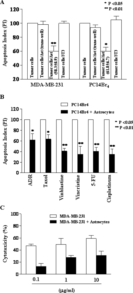
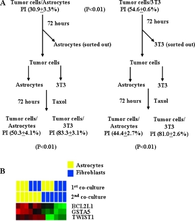
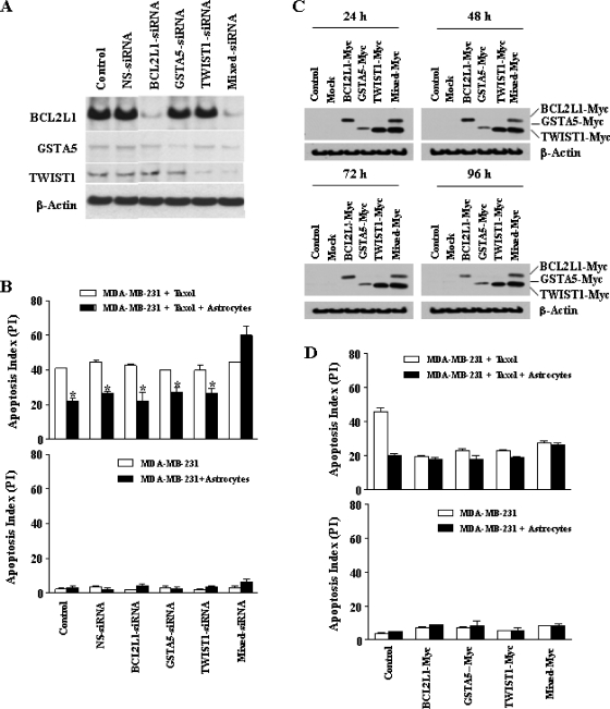
In the United States, more than 40% of cancer patients develop brain metastasis. The median survival for untreated patients is 1 to 2 months, which may be extended to 6 months with conventional radiotherapy and chemotherapy. The growth and survival of metastasis depend on the interaction of tumor cells with host factors in the organ microenvironment. Brain metastases are surrounded and infiltrated by activated astrocytes and are highly resistant to chemotherapy. We report here that coculture of human breast cancer cells or lung cancer cells with murine astrocytes (but not murine fibroblasts) led to the up-regulation of survival genes, including GSTA5, BCL2L1, and TWIST1, in the tumor cells. The degree of up-regulation directly correlated with increased resistance to all tested chemotherapeutic agents. We further show that the up-regulation of the survival genes and consequent resistance are dependent on the direct contact between the astrocytes and tumor cells through gap junctions and are therefore transient. Knocking down these genes with specific small interfering RNA rendered the tumor cells sensitive to chemotherapeutic agents. These data clearly demonstrate that host cells in the microenvironment influence the biologic behavior of tumor cells and reinforce the contention that the organ microenvironment must be taken into consideration during the design of therapy.


Figures

Figure 1
Figure 1
(A) Immunohistochemical analysis of mouse Lewis lung carcinoma (3LL) in the brain of a C57BL/6 mouse. Dividing 3LL cells (PCNA+, blue) are infiltrated and surrounded by activated astrocytes (GFAP+, brown). (B) Immunofluorescent staining of experimental brain metastasis produced by intracarotid injection of human lung adenocarcinoma PC14Br4 cells into nude mouse. Activated astrocytes were stained with GFAP polyclonal antibody (red) and nuclei were stained with SYTOX green. Tumor cells (large nuclei) are surrounded by activated astrocytes. (C) Scanning electron microscopy of in vitro culture of human breast cancer MDA-MB-231 cells (T) surrounding a murine astrocyte (A). Note astrocyte end-feet touching tumor cells.
Figure 2
Figure 2
Chemoprotection assay by PI staining and FACS analysis (A, B) or MTT assay (C). (A) Note the significant (P < .01) decrease in apoptotic index in tumor cells directly cocultured with astrocytes but not in those cocultured with fibroblasts. Separating the tumor cells from astrocytes with a 0.4-µm pore membrane prevented induction of chemoprotection. (B) Astrocyte-mediated chemoprotection against different chemotherapeutic drugs. Coculture of human lung cancer cell PC14Br4 with astrocytes induced significant protection against all P-glycoprotein-dissociated drugs. (C) Astrocyte protection of tumor cells from vinblastine measured by MTT assay. At various concentrations of vinblastine, coculture of human breast cancer cell MDA-MB-231 with astrocytes induced significant protection (P <.01).
Figure 3
Figure 3
(A) Gap junction assay. Transfer of calcein AM from the donor cells to the recipient cells was observed only when donor cells and recipient cells were cocultured in direct contact. When cells were cultured in Transwell system, which inhibited direct cell contact between donor and recipient cells, gap junction was not established between them, and consequently, calcein AM was not transferred to the donor cells. (B and C) Effect of CBX on chemoprotection. Chemoprotection assay was performed by PI staining and FACS analysis. CBX significantly reversed the protective effects induced by astrocytes in MDA-MB-231 (B) and PC14Br4 (C). *P < .01.
Figure 4
Figure 4
(A) Measured expression patterns of human and mouse genes by cross-species hybridization with human and mouse RNA microarray slides. The data are presented in matrix format in which rows represent individual gene and columns represent each hybridization event. Each cell in the matrix represents the expression level of a gene feature in an individual hybridization. The red in cells reflects measured expression levels, and intensity of color represents the magnitude of expression (log2-transformed scale). These results confirm the specificity of the analyses. (B) Expression patterns of human genes in MDA-MB-231 and PC14Br4 cocultured with murine astrocytes or 3T3 cells. We identified genes whose expression patterns were altered on interaction with astrocytes in each cell line by applying two-sample t tests (P <.001). In MDA-MB-231 and PC14Br4 cells, 1069 and 594 genes, respectively, were differentially expressed. The expression of 205 genes was altered in both cell lines. Of these, several are related to antiapoptosis and cell survival. (C) Validation of microarray by Western blot analysis. Protein was extracted from MDA-MB-231 and PC14Br4 cells after they were cultured alone or with or murine astrocytes. The expression of the antiapoptotic survival genes BCL2L1, TWIST1, and GSTA was upregulated in MDA-MB-231 and PC14Br4 cells cocultured with murine astrocytes. (D) Expression of BCL2L1, TWIST1, and GSTA5 in clinical specimen of breast cancer and lung cancer metastasis in the brain and breast cancer in the brain and lung. BCL2L1, TWIST1, and GSTA5 were highly expressed (green) on tumor cells. Nuclei were stained with 4′,6-diamidino-2-phenylindole (blue). Tumor cells in clinical specimen of breast cancer metastases to the lung were negative for expression of these genes, whereas low expression of these genes was detected in normal alveolar epithelial cells and alveolar macrophages.
Figure 5
Figure 5
Chemoprotection requires constant contact with astrocytes. (A) The apoptotic index of PC14Br4 cells cultured with astrocytes was 30.9% ± 3.3%, and with fibroblasts, it was 54.6% ± 0.6% (P > .01). Tumor cells initially cultured with astrocytes were harvested and reincubated with astrocytes or fibroblasts in the presence of taxol for another 72 hours. Tumor cells cocultured again with astrocytes maintained the relative resistance to taxol compared with tumor cells cocultured (second cycle) with fibroblasts. Tumor cells initially cultured with fibroblasts were not resistant to taxol, but if these tumor cells were then reincubated (second cycle) with astrocytes, they developed resistance compared with tumor cells cultured again with fibroblasts. (B) Gene expression of BCL2L1, GSTA5, and TWIST1 was determined in PC14Br4 cells cultured with astrocytes or fibroblasts. Similar to data shown in Figure 3, the survival genes were highly expressed in tumor cells cocultured with astrocytes but not with fibroblasts (first cycle). We harvested surviving tumor cells and cocultured them for the second cycle with either astrocytes or fibroblasts. Once again, only tumor cells cocultured with astrocytes (second cycle) expressed higher levels of BCL2L1, GSTA5, and TWIST1.
Figure 6
Figure 6
(A) Western blot analysis for validation of knockdown of target genes using siRNA. The results demonstrate specificity of the transfection with siRNA in the down-regulation of corresponding protein expression level. (B) Upper panel. Chemoprotection assay. Reversal of the protection by astrocytes was achieved only when all three genes were knocked down. Lower panel. Transfection of MDA-MD-231 human breast cancer cells with siRNA targeting BCL2L1, GSTA5, and TWIST1 did not affect the apoptosis index. (C) Stable expression of BCL2L1, GSTA5, and TWIST1 genes. Western blot analysis using anti-Myc antibody shows stable overexpression of targeted genes at the indicated time points. (D) Effects of overexpression of BCL2L1, GSTA5, and TWIST1 genes of tumor cells on chemoprotection mediated by murine astrocytes. Overexpression of BCL2L2, GSTA5, TWIST1 alone or all three genes increased tumor cell resistance to taxol in the absence of astrocytes (upper panel). Overexpression of single gene or pool of genes did not affect the apoptosis index of tumor cells in control cultures (lower panel). *P < .01.
Figure 7
Figure 7
(A) Western blot analysis for expression of AKT/pAKT and MAPK/pMAPK in human breast cancer cells and lung cancer cells cultured alone or cocultured with astrocytes or fibroblasts. Expression of AKT and MAPK was not altered whether tumor cells were cultured alone or cocultured with astrocytes or fibroblasts. However, the expression of phosphorylated AKT or MAPK was upregulated only in tumor cells cocultured with astrocytes. (B) Determination of the role of BCL2L1, GSTA5, and TWIST1 genes in AKT and MAPK activation. Transfection with nonspecific (NS-siRNA) and combined siRNA (Mixed siRNA) did not affect activation of AKT and MAPK pathways. (C) Determination of the role of AKT and MAPK activation in the regulation of BCL2L1, GSTA5, and TWIST1 gene expression. Inhibition of activation of the AKT and MAPK pathways inhibited the up-regulation of the expression of BCL2L1, GSTA5 and TWIST1 genes. All figures are representative of one experiment of three independent experiments.

References

    1. Sawaya R, Bindal R, Lang FF. Metastatic brain tumors. In: Kaye AH, Laws EE, editors. Brain Tumors. New York, NY: Churchill-Livingstone; 2001. pp. 999–1026.
    1. Landis SH, Murray T, Bolden S, Wingo PA. Cancer statistics, 1998. CA Cancer J Clin. 1998;48:6–29. - PubMed
    1. Norden AD, Wen PY, Kesari S. Brain metastases. Curr Opin Neurol. 2005;18:654–661. - PubMed
    1. Palmieri D, Smith QR, Lockman PR, Bronder J, Gril B, Chambers AF, Weil RJ, Steeg PS. Brain metastases of breast cancer. Am J Pathol. 2006;26:139–147. - PubMed
    1. Abbott N. Astrocytes-endothelial interactions and blood-brain barrier permeability. J Anat. 2002;200:629–638. - PMC - PubMed

Publication types

MeSH terms