. 2011 Feb;7(2):e1001289.
doi: 10.1371/journal.ppat.1001289. Epub 2011 Feb 17.

Secreted bacterial effectors that inhibit host protein synthesis are critical for induction of the innate immune response to virulent Legionella pneumophila

Affiliations

Secreted bacterial effectors that inhibit host protein synthesis are critical for induction of the innate immune response to virulent Legionella pneumophila

Mary F Fontana et al. PLoS Pathog. 2011 Feb.

Abstract

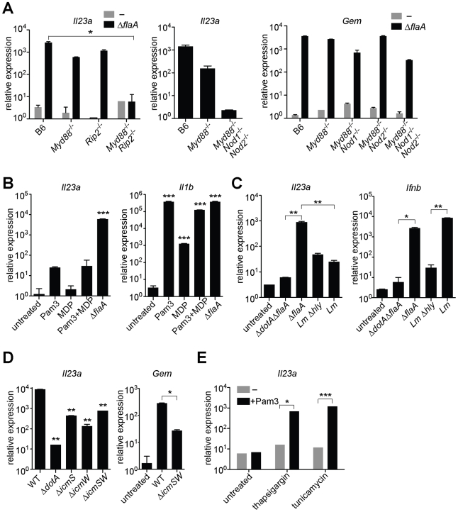
The intracellular bacterial pathogen Legionella pneumophila causes an inflammatory pneumonia called Legionnaires' Disease. For virulence, L. pneumophila requires a Dot/Icm type IV secretion system that translocates bacterial effectors to the host cytosol. L. pneumophila lacking the Dot/Icm system is recognized by Toll-like receptors (TLRs), leading to a canonical NF-κB-dependent transcriptional response. In addition, L. pneumophila expressing a functional Dot/Icm system potently induces unique transcriptional targets, including proinflammatory genes such as Il23a and Csf2. Here we demonstrate that this Dot/Icm-dependent response, which we term the effector-triggered response (ETR), requires five translocated bacterial effectors that inhibit host protein synthesis. Upon infection of macrophages with virulent L. pneumophila, these five effectors caused a global decrease in host translation, thereby preventing synthesis of IκB, an inhibitor of the NF-κB transcription factor. Thus, macrophages infected with wildtype L. pneumophila exhibited prolonged activation of NF-κB, which was associated with transcription of ETR target genes such as Il23a and Csf2. L. pneumophila mutants lacking the five effectors still activated TLRs and NF-κB, but because the mutants permitted normal IκB synthesis, NF-κB activation was more transient and was not sufficient to fully induce the ETR. L. pneumophila mutants expressing enzymatically inactive effectors were also unable to fully induce the ETR, whereas multiple compounds or bacterial toxins that inhibit host protein synthesis via distinct mechanisms recapitulated the ETR when administered with TLR ligands. Previous studies have demonstrated that the host response to bacterial infection is induced primarily by specific microbial molecules that activate TLRs or cytosolic pattern recognition receptors. Our results add to this model by providing a striking illustration of how the host immune response to a virulent pathogen can also be shaped by pathogen-encoded activities, such as inhibition of host protein synthesis.


Conflict of interest statement

The authors have declared that no competing interests exist.

Figures

Figure 1
Figure 1. A unique transcriptional response in macrophages infected with virulent L. pneumophila.
(A) Caspase-1−/− macrophages were infected for 6 h with the specified strains of L. pneumophila. RNA was amplified and hybridized to MEEBO microarrays. Black and red dots, genes exhibiting greater than 2-fold difference in induction between wildtype (WT) and mutant. Red dots indicate labeled genes. Data shown are the average of two experiments. (B) B6 macrophages were infected for 6 h with the specified strains of L. pneumophila. Levels of the indicated transcripts were measured by quantitative RT-PCR. (C) Mice were infected intranasally with 2×10⁶ L. pneumophila and bronchoalveolar lavage was performed 24 h post infection. Host cells recovered from bronchoalveolar lavage fluid (BALF) were counted with a hemocytometer. A portion of each sample was plated on BCYE plates to enumerate cfu. (D) Macrophages were infected for 6 h with L. pneumophila. Levels of the indicated transcripts were measured by quantitative RT-PCR. N.S., not significant. Data shown are representative of two (a, d) or at least three (B, C) experiments (mean ± sd in b, d). *, p<0.05 versus uninfected. ***, p<0.005 versus uninfected.
Figure 2
Figure 2. MyD88 and Nod signaling alone do not account for the unique response to virulent L. pneumophila, which can be recapitulated by ER stress inducers that also inhibit translation.
In all panels, the indicated transcripts were measured by quantitative RT-PCR. (A) Macrophages were infected with ΔflaA L. pneumophila for 6 h. (B) Macrophages were infected with L. pneumophila or were treated with Pam3CSK4 (10 ng/mL) and/or transfected with MDP (10 µg/mL) for 6 h. (C) B6 macrophages were infected with L. pneumophila, wildtype L. monocytogenes or the avirulent L. monocytogenes Δhly mutant for 4 h. (D) B6 macrophages were infected with the indicated strains of L. pneumophila for 6 h. **, p<0.01 compared to wildtype (WT). (E) Uninfected B6 macrophages were treated with thapsigargin (500 nM) or tunicamycin (5 µg/mL) for 6 h alone or in conjunction with Pam3CSK4 (1 ng/mL). All results shown are representative of at least three experiments (mean ± sd). Lm, L. monocytogenes. *, p<0.05; **, p<0.01; ***, p<0.005.
Figure 3
Figure 3. A mutant L. pneumophila lacking 5 bacterial effectors that inhibit host protein synthesis is defective in induction of the host ‘effector-triggered response’.
Growth of the indicated strains of L. pneumophila was measured in amoebae (A) or A/J macrophages (B). (C) Global host protein synthesis was measured by 35S-methionine incorporation in macrophages infected for 2.5 h with the indicated strains. (D) Myd88−/− (bottom right graph) or Caspase-1−/− (all others) macrophages were infected for 6 h with the specified strains. The indicated transcripts were measured by quantitative RT-PCR. (E) Caspase-1−/− macrophages were infected for 6 h with the specified strains. Indicated strains carried plasmids that constitutively expressed either a functional (plgt2, plgt3) or a catalytically inactive (plgt2*, plgt3*) bacterial effector. Data shown are representative of two (b, c) or at least three (A, D, E) experiments (mean ± sd). Δ5, Δlgt1Δlgt2Δlgt3ΔsidIΔsidL. Δ4, Δlgt1Δlgt2Δlgt3ΔsidI. *, p<0.05. ***, p<0.005.
Figure 4
Figure 4. Induction of the ‘effector-triggered response’ can be recapitulated by pharmacological inhibitors of translation.
(A) B6 macrophages were infected for 6 h with the indicated strains, alone or with CHX (5 µg/mL). (B, C, D) B6 macrophages were infected or were treated for 4 h with CHX (10 µg/mL; B), puromycin (20 µg/mL; C) or bruceantin (50 nM; D) alone or in conjunction with Pam3CSK4 (10 ng/mL). CHX, cycloheximide. Data shown are representative of two (C, D) or three (A, B) experiments (mean ± sd). *, p<0.05. **, p<0.01.
Figure 5
Figure 5. Expression of the 5 L. pneumophila effectors and induction of ‘effector-triggered’ genes correlates with sustained loss of inhibitors of the NF-κB transcription factor.
(A) Caspase-1−/− macrophages were infected for 6 h with the indicated strains. RNA was amplified and hybridized to MEEBO arrays. Black and red dots, genes exhibiting greater than 2-fold difference in induction between wildtype (WT) and Δ5. Red dots indicate labeled genes. (B, C) Caspase-1−/− macrophages were infected at an MOI of 2 for the times indicated. Cell lysates were analyzed by Western blotting with anti-IκBα antibody (top panels) or anti-β-actin antibody (bottom panels). (C) The indicated strains carried a plasmid encoding either a functional (plgt3) or catalytically inactive (plgt3*) effector. (D) B6 macrophages were infected at an MOI of 2 for the times indicated. Nuclear extracts were analyzed by Western blotting with anti-NF-κB antibody (top panel) or anti-lamin-B antibody (bottom panel) as a loading control. Cytoplasmic extract of untreated macrophages (CE) was included for comparison. (E, F) B6 (E, F) or A20−/− (E) macrophages were infected for 6 h, and levels of the indicated transcripts were measured by quantitative RT-PCR. Data shown are representative of two experiments (E-F, mean ± sd).
Figure 6
Figure 6. Model of NF-κB activation and superinduction by translation inhibitors.
(A) NF-κB activation by TLR signaling, via the adaptor Myd88, or Nod signaling, via Rip2, normally leads to synthesis of inhibitory proteins, including IκB and A20, which act to shut off NF-κB signaling. (B) When translation is inhibited, IκB and A20 fail to be synthesized, allowing sustained activation of NF-κB and subsequent robust transcription of a subset of target genes.
Figure 7
Figure 7. Inhibition of host translation by multiple bacterial toxins provokes an inflammatory cytokine response in vitro and in vivo.
(A) B6 macrophages were infected for 24 h with the indicated strains of L. pneumophila and/or treated with cycloheximide (5 µg/mL). Protein levels in the supernatant were assayed by ELISA. (B) B6 macrophages were treated for 5 h with Diphtheria Toxin (1 ng/mL; left panel) or with Exotoxin A (500 ng/mL; right panel), alone or in conjunction with Pam3CSK4. Il23a transcript levels were assayed by quantitative RT-PCR. n.d., not detected. (C) B6 mice were treated intranasally with Pam3CSK4 (10 µg/mouse) or ExoA (2 µg/mouse) or both in 25 µL PBS. Bronchoalveolar lavage was performed 24 h post infection. GM-CSF levels in lavage were measured by ELISA. Data are representative of two (A, C) or three (B) experiments (mean ± sd in A, B). CHX, cycloheximide. DT, Diphtheria Toxin. ExoA, Exotoxin A. *, p<0.05. ***, p<0.005.

References

    1. Takeuchi O, Akira S. Pattern recognition receptors and inflammation. Cell. 2010;140:805–820. - PubMed
    1. Rakoff-Nahoum S, Paglino J, Eslami-Varzaneh F, Edberg S, Medzhitov R. Recognition of commensal microflora by toll-like receptors is required for intestinal homeostasis. Cell. 2004;118:229–241. - PubMed
    1. Vance RE, Isberg RR, Portnoy DA. Patterns of pathogenesis: discrimination of pathogenic and nonpathogenic microbes by the innate immune system. Cell Host Microbe. 2009;6:10–21. - PMC - PubMed
    1. Medzhitov R. Innate immunity: quo vadis? Nat Immunol. 2010;11:551–553. - PubMed
    1. Chisholm ST, Coaker G, Day B, Staskawicz BJ. Host-microbe interactions: shaping the evolution of the plant immune response. Cell. 2006;124:803–814. - PubMed

Publication types

MeSH terms