. 2011 Mar;7(3):e1001097.
doi: 10.1371/journal.pcbi.1001097. Epub 2011 Mar 3.

Dynamic conformational changes in munc18 prevent syntaxin binding

Affiliations

Dynamic conformational changes in munc18 prevent syntaxin binding

Dana Bar-On et al. PLoS Comput Biol. 2011 Mar.

Abstract

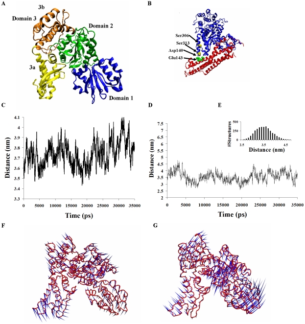
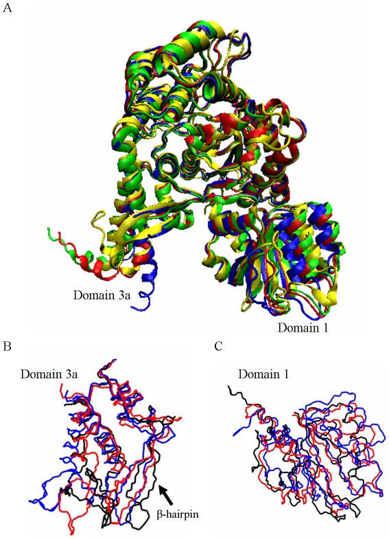
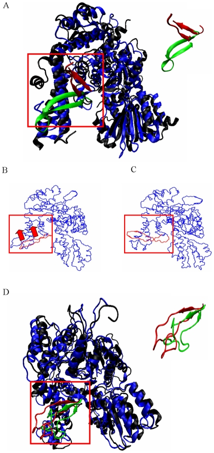
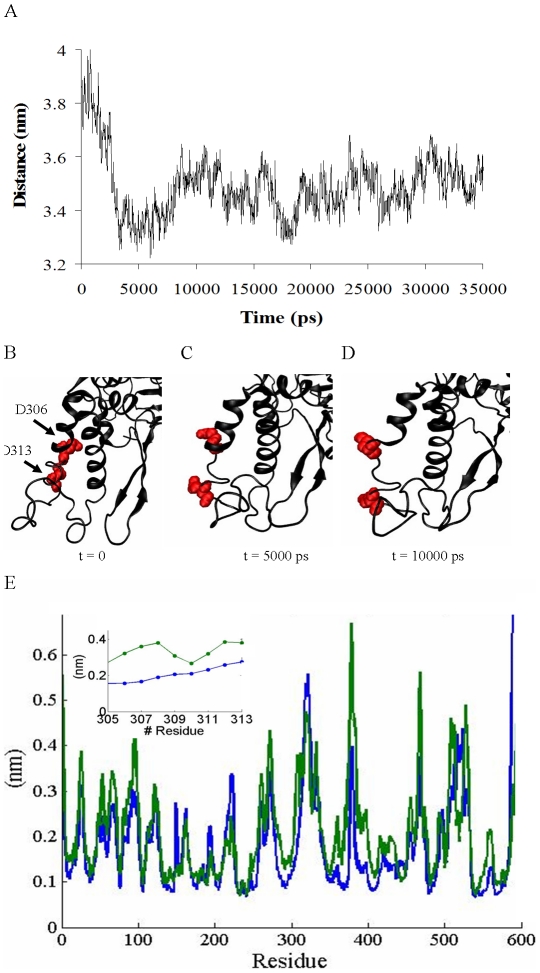
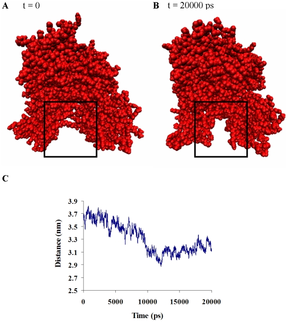
The Sec1/munc18 protein family is essential for vesicle fusion in eukaryotic cells via binding to SNARE proteins. Protein kinase C modulates these interactions by phosphorylating munc18a thereby reducing its affinity to one of the central SNARE members, syntaxin-1a. The established hypothesis is that the reduced affinity of the phosphorylated munc18a to syntaxin-1a is a result of local electrostatic repulsion between the two proteins, which interferes with their compatibility. The current study challenges this paradigm and offers a novel mechanistic explanation by revealing a syntaxin-non-binding conformation of munc18a that is induced by the phosphomimetic mutations. In the present study, using molecular dynamics simulations, we explored the dynamics of the wild-type munc18a versus phosphomimetic mutant munc18a. We focused on the structural changes that occur in the cavity between domains 3a and 1, which serves as the main syntaxin-binding site. The results of the simulations suggest that the free wild-type munc18a exhibits a dynamic equilibrium between several conformations differing in the size of its cavity (the main syntaxin-binding site). The flexibility of the cavity's size might facilitate the binding or unbinding of syntaxin. In silico insertion of phosphomimetic mutations into the munc18a structure induces the formation of a conformation where the syntaxin-binding area is rigid and blocked as a result of interactions between residues located on both sides of the cavity. Therefore, we suggest that the reduced affinity of the phosphomimetic mutant/phosphorylated munc18a is a result of the closed-cavity conformation, which makes syntaxin binding energetically and sterically unfavorable. The current study demonstrates the potential of phosphorylation, an essential biological process, to serve as a driving force for dramatic conformational changes of proteins modulating their affinity to target proteins.


Conflict of interest statement

The authors have declared that no competing interests exist.

Figures

Figure 1
Figure 1. Dynamic equilibrium between several open- and closed-cavity conformations of wild-type munc18a.
A) Structure of munc18a (3C98.pdb). B) Ribbon presentation of syntaxin-1a-munc18a complex including the location of the munc18a phosphorylation sites (Ser 306 and Ser 313) and adjacent residues of syntaxin-1a (Asp 140 and Glu 143). C) Time-dependent change in the distance between the centers of mass of domains 1 and 3a during the wild-type munc18a simulation (simulation 1). D) The distance between residues Gly 26 and Glu 273. E) Histogram of the distribution of the distance between Gly 26 and Glu 273. F–G) Porcupine plots based on ED analysis of two of the main motion vectors (the first and the fourth) of munc18a wild-type; the direction and the length of the ‘needles’ in blue indicate the direction and extent of the motion respectively. Closure motion of the cavity (F) opening motion of the cavity (G).
Figure 2
Figure 2. Comparison of free munc18a dynamics to its squid homologs' crystal structures.
A) Superposition of the backbone of four crystal structures presented in ribbon form; green, blue and red: the three resolved crystal structures of the squid sSec1: green (1EPU.pdb), blue (1FVF.pdb) and red (1FVH.pdb); yellow: the crystal structure of munc18a taken from the complex with syntaxin (, 3C98.pdb). B) Snapshots of domain 3a taken from the last frame of the first eigenvector in each of the three wild-type simulations; blue: 1, red: 2, black: 3. C) Snapshots of domain 1 taken from the last frame of the main eigenvectors of each of the three MD simulations; blue: 1, red: 2, black: 3.
Figure 3
Figure 3. Comparison of munc18a dynamics with the crystal structure of Sly1p, the yeast Golgi homolog.
A) Superposition of the munc18a crystal structure taken from its structure in the complex with syntaxin (3C98.pdb, [12]) and the crystal structure of Sly1p, the yeast Sec1/munc18 protein, taken from its crystal structure with the N-peptide of the Golgi syntaxin, Sed5p (1mqs.pdb, [23]). The proteins are presented in ribbon form, in blue and black, respectively. The β-hairpin in domain 3a, present in both of the structures, is marked in green and red, respectively. Inset: The β-hairpin in domain 3a, present in both of the structures, is marked in green and red, respectively. Note the difference in the hairpin's positions in the two structures. B, C) The first eigenvector of simulation 2 exhibits substantial movement of the β-hairpin in domain 3a upwards. B) The first frame of the eigenvector movie (see Methods), representing the starting point of the movement. The red arrows indicate the upward direction of the movement of the β-hairpin during the simulation. C) The last frame of the movie's eigenvector (see Methods), representing the maximum point of the movement. D) Superposition of the Sly1p full-length reconstructed structure at t = 0 (black) and after 15 ns of MD simulation (blue) both presented in the ribbon form. The different positions of the β-hairpin in domain 3a are presented (in red and green respectively). Inset: magnified superposition of the β-hairpin in domain 3a, present in both of the structures (green and red, respectively).
Figure 4
Figure 4. Cavity closure in munc18aS306D,S313D phosphomimetic mutant structure.
A) Measurements of the distance between the centers of mass of domains 3a and 1 during the simulation of mutant munc18aS306D,S313D. B–D) Structural changes in the positions of the phosphomimetic residues (Glu 306 and Glu 313) of munc18a (simulation M1, Table 1). Snapshots taken from the MD simulation of munc18aS306D,S313D; at B) t = 0, C) t = 5000 ps, D) t = 10000 ps. E) Comparison of the RMSF values of munc18a residues in the wild-type (simulation 1) versus the phosphomimetic mutant simulations (blue and green curves, respectively). Inset: magnification of E in the area of the phosphomimetic mutations (residues 305–313), exemplifying the higher RMSF values in this region in the munc18aS306D,S313D simulation.
Figure 5
Figure 5. The closure of the cavity as detected by ED analyses of munc18aS306D, S313D simulation.
A) Porcupine plot, presenting the first eigenvector in the mutant munc18aS306D,S313D simulation as produced by ED analysis (M1) performed by the Dynatraj tool. B) Comparison of the magnitudes of the eight main eigenvectors of the GROMACS-based ED analysis extracted from the simulations of wild-type munc18a (simulation 1, blue squares) and munc18aS306D,S313D (simulation M1, green squares). C, D) Snapshots of the first (t = 0, C) and the last (t = 35 ns, D) frames of the second munc18aS306D,S313D (Simulation M2, Table 1) simulation.
Figure 6
Figure 6. Energetic stabilization is correlated to munc18aS306D,S313D closure of the cavity.
A) Averaged total energy changes during the simulation of the munc18aS306D,S313D (simulation M1). B) Time-dependent changes in the Coulomb energy component. C) Time-dependent change in the number of hydrogen bonds between domains 3a and 1 in the simulation of the wild-type munc18a. D) Hydrogen-bond formation between domains 3a and 1 during the simulation of the munc18aS306D,S313D (M1).
Figure 7
Figure 7. Electrostatic and LJ interactions of residues on either side of the munc18a cavity stabilize its closure.
A, B) Snapshots taken from the mutant munc18aS306D,S313D simulation; Munc18aS306D,S313D charged residues are presented in the space-fill model (blue, positive residues and red, negative residues). A) t = 0, B) t = ∼37 ns. Hydrophobic interactions further stabilize the closure of the cavity; hydrophobic residues in domain 1 (green) and in domain 3a (yellow). C) The distance between Lys 46 and Asp 262 during the ∼37-ns simulation (dt = 10 ps). D, E) snapshots taken from the simulation showing the position of Lys 46 and Asp 262 at D) t = 0, E) t = ∼37 ns.
Figure 8
Figure 8. Back-mutations in munc18a restore its dynamic nature and induce gradual reopening of its cavity.
A) Measurement of the distance between the centers of mass of domains 3a and 1 during back-mutated wild-type munc18a simulation (munc18aD306S,D313S, 36 ns). B) The distance of the centers of mass of two regions adjacent to the cavity: residues 35–70 (domain 1) and residues 260–280 (domain 3a). C) Porcupine plot demonstrating the opening motion of the cavity (the fourth most dominant eigenvector of the protein in the simulation). D) Superposition of domains 3a and 1 from the last frames in the simulations of munc18aS306D,S313D (t = 35 ns, red) and munc18aD306S,D313S (t = 36 ns, blue). E) Measurement of the distance between the centers of mass of domains 3a and 1 during the munc18aS306A,S313A simulation (36 ns). F) Time-dependent change in the number of hydrogen bonds between domains 3a and 1 in the munc18aS306A,S313A simulation G) Superposition of the structure of munc18aS306A,S313A in the first (t = 0, red) and last frame (t = 36 ns, blue) of the simulation.
Figure 9
Figure 9. Munc18aF115E adopts a closed-cavity conformation.
Two snapshots taken from a 20-ns-simulation of munc18aF115E demonstrate the closure of the cavity between domains 3a and 1 during the simulation of this mutant as well. A) t = 0, B) t = 20 ns, the cavity area is framed in both A and B. C) Measurement of the distance between the centers of mass of domains 3a and 1 during the Munc18aF115E simulation.

References

    1. Sudhof TC, Rothman JE. Membrane fusion: grappling with SNARE and SM proteins. Science. 2009;323:474–477. - PMC - PubMed
    1. Garcia EP, Gatti E, Butler M, Burton J, De Camilli P. A rat brain Sec1 homologue related to Rop and UNC18 interacts with syntaxin. Proc Natl Acad Sci U S A. 1994;91:2003–2007. - PMC - PubMed
    1. Hata Y, Slaughter CA, Sudhof TC. Synaptic vesicle fusion complex contains unc-18 homologue bound to syntaxin. Nature. 1993;366:347–351. - PubMed
    1. Gengyo-Ando K, Kamiya Y, Yamakawa A, Kodaira K, Nishiwaki K, et al. The C. elegans unc-18 gene encodes a protein expressed in motor neurons. Neuron. 1993;11:703–711. - PubMed
    1. Voets T, Toonen RF, Brian EC, de Wit H, Moser T, et al. Munc18-1 promotes large dense-core vesicle docking. Neuron. 2001;31:581–591. - PubMed

Publication types