. 2011 Feb;7(2):e1001084.
doi: 10.1371/journal.pcbi.1001084. Epub 2011 Feb 24.

Dynamically-driven inactivation of the catalytic machinery of the SARS 3C-like protease by the N214A mutation on the extra domain

Affiliations

Dynamically-driven inactivation of the catalytic machinery of the SARS 3C-like protease by the N214A mutation on the extra domain

Jiahai Shi et al. PLoS Comput Biol. 2011 Feb.

Abstract

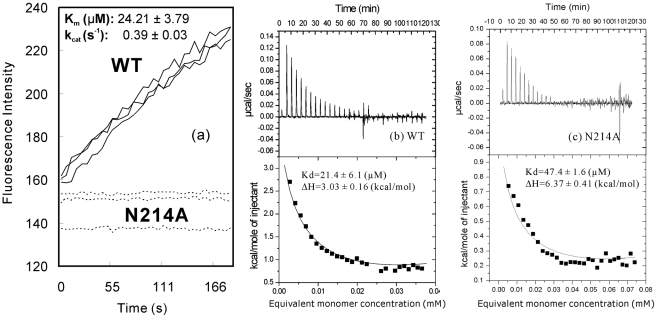
Despite utilizing the same chymotrypsin fold to host the catalytic machinery, coronavirus 3C-like proteases (3CLpro) noticeably differ from picornavirus 3C proteases in acquiring an extra helical domain in evolution. Previously, the extra domain was demonstrated to regulate the catalysis of the SARS-CoV 3CLpro by controlling its dimerization. Here, we studied N214A, another mutant with only a doubled dissociation constant but significantly abolished activity. Unexpectedly, N214A still adopts the dimeric structure almost identical to that of the wild-type (WT) enzyme. Thus, we conducted 30-ns molecular dynamics (MD) simulations for N214A, WT, and R298A which we previously characterized to be a monomer with the collapsed catalytic machinery. Remarkably, three proteases display distinctive dynamical behaviors. While in WT, the catalytic machinery stably retains in the activated state; in R298A it remains largely collapsed in the inactivated state, thus implying that two states are not only structurally very distinguishable but also dynamically well separated. Surprisingly, in N214A the catalytic dyad becomes dynamically unstable and many residues constituting the catalytic machinery jump to sample the conformations highly resembling those of R298A. Therefore, the N214A mutation appears to trigger the dramatic change of the enzyme dynamics in the context of the dimeric form which ultimately inactivates the catalytic machinery. The present MD simulations represent the longest reported so far for the SARS-CoV 3CLpro, unveiling that its catalysis is critically dependent on the dynamics, which can be amazingly modulated by the extra domain. Consequently, mediating the dynamics may offer a potential avenue to inhibit the SARS-CoV 3CLpro.


Conflict of interest statement

The authors have declared that no competing interests exist.

Figures

Figure 1
Figure 1. Enzymatic activity and dissociation constant of the dimer-monomer equilibrium.
(a). Enzymatic activities of the WT (black lines) and N214A (dotted lines) proteases by monitoring the increase of the emission fluorescence intensity at a wavelength of 538 nm continuously for 3 min. The Km and kcat values are presented for the WT enzyme. The ITC dilution profiles for measuring the dissociation constants of the dimer-monomer equilibrium for WT (b) and N214A (c). The Kd and ΔH values were obtained by fitting the ITC data with the built-in Microcal ORIGIN software.
Figure 2
Figure 2. Crystal structure of the N214A mutant.
(a). Overall superimposition of the dimeric N214A (violet) and WT (cyan; PDB code of 2H2Z) structures. (b) Dimeric structure of the SARS 3CLpro showing the catalytic dyad His41-Cys145 located on the cleft of domain I (red) and domain II (blue) of the chymotrypsin fold, as well as Asn214 on the extra domain. Superimpositions of the catalytically critical residues of WT (violet) and N214A (cyan; PDB code of 2H2Z) for protomers A (c) and B (d) respectively.
Figure 3
Figure 3. Overall dynamic behaviors in the MD simulations.
Root-mean-square deviations (RMSD) of the heavy atoms for three independent MD simulations of the WT protomer A (black) and B (red) (a–c); R298A (black) (d–e); N214A protomer A (black) and B (red) (g–i). Root-mean-square fluctuations of the Cα atoms computed for three independent simulations for WT (j–l), R298A (m–o) and N214A (p–r).
Figure 4
Figure 4. Structure snapshots in the MD simulations.
The conformations of the residues constituting the catalytic machinery at 0 ns (yellow), 10 ns (cyan), 20 ns (blue) and 30 ns (red) for three independent simulations of WT, R298A and N214A respectively.
Figure 5
Figure 5. Dynamic behavior of the catalytic dyad.
Time-trajectories of the distance between NE2 of His41 and SG of Cys145 atoms of WT (a–c); R298A (d–f) and N214A (g–i) in the 30-ns simulations. Time-trajectories of the Chi1 dihedral angle of His41 of WT (j–l); R298A (m–o) and N214A (p–r). Trajectories of the Chi2 dihedral angle of His41 of WT (s–u); R298A (v–x) and N214A (y–aa).
Figure 6
Figure 6. Dynamic behavior of the oxyanion loop residues.
Ramachandran plots of the residues Ser139-Phe140-Leu141 for WT (a–c, j–l and s–u); R298A (d–f, m–o and v–x) and N214A (g–i, p–r and y–aa). The spots are colored in black for protomer A and red for protomer B.
Figure 7
Figure 7. Dynamic behavior of the His172-Glu166 interaction.
Time-trajectories of the distances between the aromatic rings of His172 and Glu166 of WT (a–c); R298A (d–f) and N214A (g–i) in three simulations. Time-trajectories of the Chi1 dihedral angle of Glu166 of WT (j–l); R298A (m–o) and N214A (p–r). Time-trajectories of the Chi2 dihedral angle of Glu166 of WT (s–u); R298A (v–x) and N214A (y–aa).
Figure 8
Figure 8. Dynamic behavior of the Phe140-His172 interaction.
Time-trajectories of the centroid distances between the aromatic rings of Phe140 and His172 of WT (a–c); R298A (d–f) and N214A (g–i) in three independent simulations. Time-trajectories of the Chi1 dihedral angle of His172 of WT (j–l); R298A (m–o) and N214A (p–r). Time-trajectories of the Chi2 dihedral angle of His172 of WT (s–u); R298A (v–x) and N214A (y–aa).
Figure 9
Figure 9. Dynamic behavior of the mutation site.
Ramachandran plots of the residues Asn214 for WT (a–c); R298A (d–f) and Ala214 of N214A (g–i) in the 30 ns simulations. Time-trajectories of the Phi dihedral angle of Asn214 of WT (j–l); R298A (m–o), and Ala214 for N214A (p–r). Time-trajectories of the Psi dihedral angle of Asn214 of WT (s–u); R298A (v–x), and Ala214 for N214A (y–aa).

References

    1. Anand K, Palm GJ, Mesters JR, Siddell SG, Ziebuhr J, et al. Structure of coronavirus main proteinase reveals combination of a chymotrypsin fold with an extra alpha-helical domain. EMBO J. 2002;21:3213–24. - PMC - PubMed
    1. Anand K, Ziebuhr J, Wadhwani P, Mesters JR, Hilgenfeld R. Coronavirus main proteinase (3CLpro) structure: basis for design of anti- SARS drugs. Science. 2003;300:1763–1767. - PubMed
    1. Allaire M, Chernaia MM, Malcolm BA, James MN. Picornaviral 3C cysteine proteinases have a fold similar to chymotrypsin-like serine proteinases. Nature. 1994;369:72–6. - PubMed
    1. Yang H, Yang M, Ding Y, Liu Y, Lou Z, et al. The crystal structures of severe acute respiratory syndrome virus main protease and its complex with an inhibitor. Proc Natl Acad Sci U S A. 2003;100:13190–5. - PMC - PubMed
    1. Barrila J, Bacha U, Freire E. Long-range cooperative interactions modulate dimerization in SARS 3CLpro. Biochemistry. 2006;45:14908–16. - PMC - PubMed

Publication types

MeSH terms