. 2011 Feb 24;6(2):e16972.
doi: 10.1371/journal.pone.0016972.

Critical role of neuropeptides B/W receptor 1 signaling in social behavior and fear memory

Affiliations

Critical role of neuropeptides B/W receptor 1 signaling in social behavior and fear memory

Ruby Nagata-Kuroiwa et al. PLoS One. .

Abstract

Neuropeptide B/W receptor 1 (NPBWR1) is a G-protein coupled receptor, which was initially reported as an orphan receptor, and whose ligands were identified by this and other groups in 2002 and 2003. To examine the physiological roles of NPBWR1, we examined phenotype of Npbwr1⁻/⁻ mice. When presented with an intruder mouse, Npbwr1⁻/⁻ mice showed impulsive contact with the strange mice, produced more intense approaches toward them, and had longer contact and chasing time along with greater and sustained elevation of heart rate and blood pressure compared to wild type mice. Npbwr1⁻/⁻ mice also showed increased autonomic and neuroendocrine responses to physical stress, suggesting that impairment of NPBWR1 leads to stress vulnerability. We also observed that these mice show abnormality in the contextual fear conditioning test. These data suggest that NPBWR1 plays a critical role in limbic system function and stress responses. Histological and electrophysiological studies showed that NPBWR1 acts as an inhibitory regulator on a subpopulation of GABAergic neurons in the lateral division of the CeA and terminates stress responses. These findings suggest important roles of NPBWR1 in regulating amygdala function during physical and social stress.


Conflict of interest statement

Competing Interests: The authors have declared that no competing interests exist.

Figures

Figure 1
Figure 1. Increased impulsiveness and contact time with associated increased autonomic responses in Npbwr1 / mice during resident-intruder test.
(A) Male naive 8-week-old mice were housed individually for 4 weeks before the procedure. The behavior of mice was recorded with a CCD video camera. A randomly chosen male intruder (C57BL/6J) was used only once in each session. The intruder was introduced into the resident cage, and behavior was recorded for 10 min. A variety of social behaviors were scored including the latency to the first aggressive contact (left panel) and time spent in aggressive contact (sniffing, rattling, chasing, mounting, wrestling and fighting) (right panel). Npbwr1 / mice showed a shorter latency time to contact with the intruder (F1,12 = 5.304, p = 0.040), and longer physical contact with the intruder compared with wild type mice (F1,12 = 6.068, p = 0.030). Data are presented as mean ± SEM (WT n = 6, KO n = 8). Also see movies S1 and S2, which show typical examples of behavior observed during this test. (B) Video tracking system shows traces of intruder (white) and resident (green) during 10 min session of resident-intruder test, showing that Npbwr1 / mice exhibited more sustained and insistent contact and chasing behavior. Note that the trace of Npbwr1 / mice is very similar to that of the intruder, reflecting the insistent chasing. (C) Locomotor and cardiovascular responses during resident-intruder test in radiotelemetry-implanted freely moving mice. Activity (upper panels), heart rate (HR; middle panels) and mean arterial pressure (MAP; lower panels) of resident mice (Npbwr1 / or wild type littermates) during the time course of the resident-intruder test are shown. Intruders (male C57BL/6J mice) were put in the cages at 0 min. Horizontal solid bar indicates the presence of an intruder. Baseline values were defined as the average values of parameters obtained during 10 min immediately prior to the resident-intruder test. Data are presented as mean ± SEM (wild type; n = 4, Npbwr1 /; n = 5) (*p<0.05, **p<0.01, compared to wild-type). (D) Real time PCR analysis showed that Neuropeptide B (NPB) and Neuropeptide W (NPW) mRNAs in whole brain were upregulated after the resident-intruder test for 60 min. Each level of expression was normalized by the level of Gapdh mRNA (wild type; n = 45, Npbwr1 /; n = 5).
Figure 2
Figure 2. Increased autonomic, neuroendocrine and behavioral responses to physical stress in Npbwr1 / mice.
(A) Npbwr1 / mice showed a greater increase in body temperature during repetitive handling stress (mild restriction and insertion of a probe into the rectum). (B) Corticotropin-releasing hormone (Crh) mRNA level in the hypothalamus was higher in Npbwr1 / mice than in wild type mice (left panel) (wild type; n = 7, Npbwr1 /; n = 6, F1,11 = 6.928, p = 0.023). Basal serum corticosterone level in Npbwr1 / mice was comparable to that in wild type mice (wild type; n = 12, Npbwr1 /; n = 17, F1,27 = 0.700, p = 0.410), but these mice showed a greater increase in corticosterone after 10 minutes of restraint stress (right panel) (wild type; n = 3, Npbwr1 /; n = 3, F1,4 = 30.732, p = 0.005). (C) Npbwr1 / mice did not show overt anxiety in the basal state, but they show increased impulsiveness to environmental challenges. Left panel, open-field test. Percentage of time spent in the center was not significantly different between Npbwr1 / mice and wild type mice (wild type; n = 23, Npbwr1 /; n = 17, F1,38 = 0.551, p = 0.463). Middle panel, elevated-plus maze test. Number of entries into open arms and time spent in open arms during 5 min test session were not different between genotypes (wild type; n = 20, Npbwr1 /; n = 24, F1,42 = 1.734, p = 0.195 and F1,42 = 2.089, p = 0.156, respectively). Right panel, light-dark exploration test. The total number of transitions, time spent in the light side, and latency until mice escaped to the dark side were recorded for 10 min after a single mouse was placed in the light compartment. Latency to enter the dark chamber from the light chamber is significantly shorter in Npbwr1 / mice (wild type; n = 17, Npbwr1 /; n = 17, F1,32 = 10.136, p = 0.003). Data are presented as mean ± SEM.
Figure 3
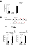
Figure 3. Npbwr1 / mice showed abnormality in contextual fear conditioning.
(A) Fear conditioning was performed to examine the ability of Npbwr1 / mice to learn and remember an auditory cue or context that predicted electric shock. Bars show the mean percentage of time spent freezing (defensive tonic immobility) during 30 s observation. For contextual fear test, mice were tested in the absence of cues in the same context at 24 hr after training. For cued test, mice were tested in new cages and the auditory cue applied. Freezing behavior of mice was counted before (pre-CS) and during application of the cue. There was a significant difference in duration of freezing behavior between Npbwr1 / mice and wild type mice during the contextual fear task (wild type; n = 13, Npbwr1 /; n = 18, F1,29 = 114.15, p<0.001), while no significant difference in freezing behavior was observed during auditory-cued testing under the altered context. (B) Alternative protocols for fear and safety conditioning . Upper panel, schematic representation of training and testing protocols. Mice were put in the conditioning chamber for 2 min before the first stimulus. Conditioning sessions consisted of 5 CS (20 s) (interval, mean 130 s, range, 100–140 s). In safety conditioning sessions, the US was explicitly unpaired and occurred during the inter-CS interval (five US per session, separated by 20–80 s from each CS). Training sessions were conducted for three days (one session per day). In fear conditioning, the US was applied for the last two sec of the CS, which was applied at the same protocol as the safety conditioning. In the test sessions, CS was delivered at the same protocol as conditioning, and no US was delivered. Freezing times in the 20 s periods before and during CS application were scored as context and cued conditioning, respectively. Lower panels, Times spent freezing during 20 s CS and 20 s prior to CS in safety conditioning (left panel) and fear conditioning (right panel) are shown. In safety conditioning, wild type mice displayed freezing to the context, which invariably accrued with US exposure. The freezing was reduced by the arrival of the CS. However, Npbwr1 / mice did not show fear responses to the context, and exhibited freezing to the CS. In fear conditioning, we obtained virtually the same results as those with the classical protocol (A).
Figure 4
Figure 4. Function of NPBWR1 in regulation of CeA neurons.
(A) Left panel, Dual-label In situ hybridization histochemistry showed co-localization of Npbwr1 mRNA (blue) with Gad67-expressing neurons (red) in the CeAl of mice. Scale bar equals 250 µm. Middle panel, higher power view of yellow rectangle region in the left panel. Right panel, high power view of yellow rectangle region in the middle panel. Opt, optic tract. (B) Left panels, typical examples of whole cell patch-clamp recording from GAD67-expressing neurons in Gad67-gfp brain sections, showing that bath-application of NPB (upper panel, 500 nM) or NPW (lower panel, 500 nM) potently inhibited neuronal activity. Right panel, numbers of GFP-positive neurons activated or inhibited by NPB/W application. We did not observe any effects in neurons of Npbwr1 / mice. (C) A typical example of morphology of NPB/W-inhibited GABAergic neurons as revealed by neurobiotin injection after patch-clamp recordings. This cell resides in the medial region of the CeAl and sends long projections to outside of the amygdala. (D) Schematic drawings of axonal projections of NPW and/or NPB-inhibited neurons in the CeAl. Left panel shows three neurons depicted in different colors that send axons within the CeAl. Right panel shows four neurons that send axons outside of the CeA.
Figure 5
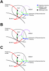
Figure 5. Schematic model of regulatory mechanism by which neuropeptide B/W regulates activity of amygdala neurons.
(A) NPB or NPW acts on NPBWR1 expressed on projection neurons in the CeAl, which could signal to the brain stem and BST to elicit emotion-related autonomic and neuroendocrine responses. Some GABAergic interneurons in the CeAl also express Npbwr1. Therefore, NPB/W signaling could modulate amygdala function in multiple pathways. (B) When the NPB/W system is activated, some of the projection neurons in the CeAl might be inhibited, while other projection neurons might be disinhibited through inhibition of GABAergic interneurons. For example, output to autonomic/neuroendocrine pathways could be inhibited, while behavioral output might be activated. (C) NPB/W system dysfunction may result in exaggerated autonomic/neuroendocrine responses along with impaired behavioral response.

References

    1. Brezillon S, Lannoy V, Franssen JD, Le Poul E, Dupriez V, et al. Identification of natural ligands for the orphan G protein-coupled receptors GPR7 and GPR8. J Biol Chem. 2003;278:776–783. Epub 2002 Oct 2024. - PubMed
    1. Fujii R, Yoshida H, Fukusumi S, Habata Y, Hosoya M, et al. Identification of a neuropeptide modified with bromine as an endogenous ligand for GPR7. J Biol Chem. 2002;277:34010–34016 Epub 32002 Jul 34012. - PubMed
    1. Tanaka H, Yoshida T, Miyamoto N, Motoike T, Kurosu H, et al. Characterization of a family of endogenous neuropeptide ligands for the G protein-coupled receptors GPR7 and GPR8. Proc Natl Acad Sci U S A. 2003;100:6251–6256 Epub 2003 Apr 6228. - PMC - PubMed
    1. O'Dowd BF, Scheideler MA, Nguyen T, Cheng R, Rasmussen JS, et al. The cloning and chromosomal mapping of two novel human opioid-somatostatin-like receptor genes, GPR7 and GPR8, expressed in discrete areas of the brain. Genomics. 1995;28:84–91. - PubMed
    1. Hondo M, Ishii M, Sakurai T. The NPB/NPW neuropeptide system and its role in regulating energy homeostasis, pain, and emotion. Results Probl Cell Differ. 2008;46:239–256. - PubMed

Publication types

MeSH terms

Substances