. 2011 Feb 24;6(2):e17285.
doi: 10.1371/journal.pone.0017285.

The minimal proteome in the reduced mitochondrion of the parasitic protist Giardia intestinalis

Affiliations

The minimal proteome in the reduced mitochondrion of the parasitic protist Giardia intestinalis

Petr L Jedelský et al. PLoS One. .

Abstract

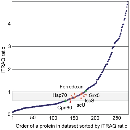
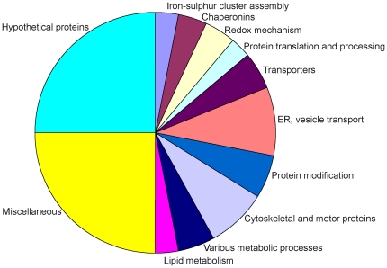
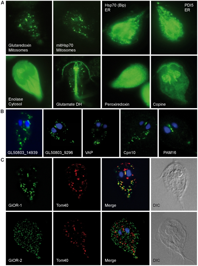
The mitosomes of Giardia intestinalis are thought to be mitochondria highly-reduced in response to the oxygen-poor niche. We performed a quantitative proteomic assessment of Giardia mitosomes to increase understanding of the function and evolutionary origin of these enigmatic organelles. Mitosome-enriched fractions were obtained from cell homogenate using Optiprep gradient centrifugation. To distinguish mitosomal proteins from contamination, we used a quantitative shot-gun strategy based on isobaric tagging of peptides with iTRAQ and tandem mass spectrometry. Altogether, 638 proteins were identified in mitosome-enriched fractions. Of these, 139 proteins had iTRAQ ratio similar to that of the six known mitosomal markers. Proteins were selected for expression in Giardia to verify their cellular localizations and the mitosomal localization of 20 proteins was confirmed. These proteins include nine components of the FeS cluster assembly machinery, a novel diflavo-protein with NADPH reductase activity, a novel VAMP-associated protein, and a key component of the outer membrane protein translocase. None of the novel mitosomal proteins was predicted by previous genome analyses. The small proteome of the Giardia mitosome reflects the reduction in mitochondrial metabolism, which is limited to the FeS cluster assembly pathway, and a simplicity in the protein import pathway required for organelle biogenesis.


Conflict of interest statement

Competing Interests: The authors have declared that no competing interests exist.

Figures

Figure 1
Figure 1. Isolation of mitosome-rich fractions.
(A) Trophozoites were disrupted and centrifuged to remove unbroken cells, nuclei and cytoskeletal residue. The high-speed pellet was resuspended in sucrose buffer, layered onto an Optiprep density gradient, and centrifuged overnight. Five distinct fractions were obtained. (B) Fractions were collected and analyzed by SDS-PAGE and Western blot. The mitosomal marker GiIscU was detected in fractions #3 and #4 using a polyclonal rabbit antibody. (C–D) Electron microscopy of subcellular fractions. Fraction #3 (C) contains numerous vesicles of variable sizes, while fraction #4 (D) contains vesicles of more homogeneous sizes. Arrows indicate mitosomes.
Figure 2
Figure 2. iTRAQ ratios define protein subcellular localization.
Proteins in fractions #3 and #4 isolated on the Optiprep gradient were labeled with the iTRAQ-114 and iTRAQ-115 reagents, respectively, analyzed by LC MS/MS, and sorted according to the iTRAQ ratios. Mitosomal marker proteins (red diamonds) fall into a narrow range of iTRAQ ratios. Green diamondsdindicate the zone of proteins considered as mitosomal candidates (mitosomal distribution, MiD).
Figure 3
Figure 3. Classification of the identified proteins according to function.
Functions were assigned based upon GiardiaDB annotations, PSI-BLAST analysis and searches of the Pfam database (Tables 1, 2, 3, 4, 5, 6 and 7, Tables S2–S3).
Figure 4
Figure 4. Sub-cellular localization of selected proteins in Giardia.
Transformed G. intestinalis cells with episomally-expressed HA-tagged proteins. (A) Marker proteins were stained using a mouse anti-HA antibody (green). Grx5, glutaredoxin 5; ER, endoplasmic reticulum; glutamate DH, glutamate dehydrogenase. (B) Predicted mitosomal proteins (GL50803_14939, GL50803_9296, VAP, Cpn10, Pam16) were stained using a mouse anti-HA antibody (green). (C) Cellular localization of tagged diflavin proteins GiOR-1 and GiOR-2 stained with mouse anti HA antibody (green). Tom40 was detected by polyclonal rabbit anti-Tom40 antibody (red).
Figure 5
Figure 5. Schematic representation of protein import pathway in the mitosome of G. intestinalis.
Components identified in mitosome are highlighted by color. Components that are known to participate in the protein import into mitochondria of animals and fungi are shown in grey colour. OM, outer membrane; IMS, intermembrane space; IM, inner membrane; TOM, translocase of outer membrane; SAM, sorting and assembly machinery; TIM, translocase of inner membrane; PAM, presequence translocase-associated motor; VAP, VAMP (vesicle-associated membrane protein)-associated protein; VDAC, voltage-dependent anion channel; MPP, mitochondrial processing peptidase

References

    1. Andersson SG, Zomorodipour A, Andersson JO, Sicheritz-Ponten T, Alsmark UC, et al. The genome sequence of Rickettsia prowazekii and the origin of mitochondria. Nature. 1998;396:133–140. - PubMed
    1. Lang BF, Burger G, O'Kelly CJ, Cedergren R, Golding GB, et al. An ancestral mitochondrial DNA resembling a eubacterial genome in miniature. Nature. 1997;387:493–497. - PubMed
    1. Vaidya AB, Mather MW. Mitochondrial evolution and functions in malaria parasites. Annu Rev Microbiol. 2009;63:249–267. 10.1146/annurev.micro.091208.073424 [doi] - PubMed
    1. Gabaldon T, Huynen MA. Shaping the mitochondrial proteome. Biochim Biophys Acta. 2004;1659:212–220. - PubMed
    1. Gabaldon T, Huynen MA. From endosymbiont to host-controlled organelle: the hijacking of mitochondrial protein synthesis and metabolism. PLoS Comput Biol. 2007;3:e219. - PMC - PubMed

Publication types