. 2012 Jan;33(1):50-62.
doi: 10.1002/hbm.21192. Epub 2011 Mar 9.

Head injury or head motion? Assessment and quantification of motion artifacts in diffusion tensor imaging studies

Affiliations

Head injury or head motion? Assessment and quantification of motion artifacts in diffusion tensor imaging studies

Josef Ling et al. Hum Brain Mapp. 2012 Jan.

Abstract

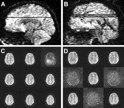
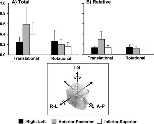
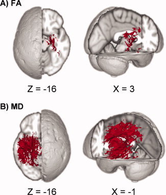
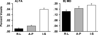
The relationship between head motion and diffusion values such as fractional anisotropy (FA) and mean diffusivity (MD) is currently not well understood. Simulation studies suggest that head motion may introduce either a positive or negative bias, but this has not been quantified in clinical studies. Moreover, alternative measures for removing bias as result of head motion, such as the removal of problematic gradients, has been suggested but not carefully evaluated. The current study examined the impact of head motion on FA and MD across three common pipelines (tract-based spatial statistics, voxelwise, and region of interest analyses) and determined the impact of removing diffusion weighted images. Our findings from a large cohort of healthy controls indicate that while head motion was associated with a positive bias for both FA and MD, the effect was greater for MD. The positive bias was observed across all three analysis pipelines and was present following established protocols for data processing, suggesting that current techniques (i.e., correction of both image and gradient table) for removing motion bias are likely insufficient. However, the removal of images with gross artifacts did not fundamentally change the relationship between motion and DTI scalar values. In addition, Monte Carlo simulations suggested that the random removal of images increases the bias and reduces the precision of both FA and MD. Finally, we provide an example of how head motion can be quantified across different neuropsychiatric populations, which should be implemented as part of any diffusion tensor imaging quality assurance protocol.


Figures

Figure 1
Figure 1
Examples of gross artifacts present in diffusion weighted (DW) images. A depicts a DW image with a single slice with signal loss whereas B depicts a DW image with multiple affected slices. C and D represent nine sequential 2 mm axial slices from an area corresponding to the artifacts displayed in A and B.
Figure 2
Figure 2
A quantitative representation of both total (A) and relative (B) motion across all healthy control subjects. For both indices, translational and rotational motion are stratified according to whether motion occurred in the right‐left (R‐L, black bars), anterior‐posterior (A‐P, grey bars), or inferior‐superior (I‐S, white bars) directions. Units for the graphs are represented in millimeters for translational motion and degrees for rotational motion. Error bars represent one standard deviation.
Figure 3
Figure 3
Results from the tract‐based spatial statistics (TBSS) analyses depicting the voxels that exhibited a significant association between motion and DTI scalar values. Data are presented for the analyses involving both fractional anisotropy (FA; A) and mean diffusivity (MD; B) as the dependent measure. Across both analyses, subjects with higher translational motion along the anterior‐posterior axis exhibited higher values of either FA or MD.
Figure 4
Figure 4
A depiction of how motion in the A‐P axis affected fibers oriented in the right‐left (R‐L, black bars), anterior‐posterior (A‐P, grey bars), or inferior‐superior (I‐S, white bars) direction during the tract‐based spatial statistics analyses. A represents a percentage of significant voxels from the fractional anisotropy analyses (FA) and B represents the voxels from the mean diffusivity (MD) analysis. Error bars represent one standard deviation.
Figure 5
Figure 5
Effects of randomly removing diffusion weighted (DW) images on both fractional anisotropy (FA) and mean diffusivity (MD) across two different subjects (please see Supp. Info., Fig. 3 for third subject). DW images were randomly selected and removed in multiples of 3 (e.g., three DW images, six DW images, etc.) before the calculation of the diffusion tensor, with each step being repeated 30 times. A presents results of a histogram analysis for all white matter voxels. The circles correspond to the mean scalar value and the error bars represent one standard deviation derived from all 30 iterations. The number of gradients removed is indicated along the x‐axis. For both subjects, a positive bias existed in the FA data associated with increasing the number of gradients removed. A positive bias was also present for two of three subjects for MD (see Supp. Info., Fig. 4), with Subject 2 exhibiting a relatively stable mean. B presents the variation in scalar values on a voxelwise basis following the random removal of gradients. For all subjects, voxelwise precision in the calculation of DTI scalar metrics decreased as a function of randomly removing DW images.

References

    1. Aksoy M, Liu C, Moseley ME, Bammer R ( 2008): Single‐step nonlinear diffusion tensor estimation in the presence of microscopic and macroscopic motion. Magn Reson Med 59: 1138–1150. - PMC - PubMed
    1. Andersson JL, Skare S ( 2002): A model‐based method for retrospective correction of geometric distortions in diffusion‐weighted EPI. Neuroimage 16: 177–199. - PubMed
    1. Ardekani BA, Tabesh A, Sevy S, Robinson DG, Bilder RM, Szeszko PR ( 2010): Diffusion tensor imaging reliably differentiates patients with schizophrenia from healthy volunteers. Hum Brain Mapp (in press). [Epub ahead of print 2010 Mar 4]. - PMC - PubMed
    1. Bazarian JJ, Zhong J, Blyth B, Zhu T, Kavcic V, Peterson D ( 2007): Diffusion tensor imaging detects clinically important axonal damage after mild traumatic brain injury: A pilot study. J Neurotrauma 24: 1447–1459. - PubMed
    1. Beaulieu C ( 2002): The basis of anisotropic water diffusion in the nervous system—A technical review. NMR Biomed 15: 435–455. - PubMed

Publication types

LinkOut - more resources