. 2012 Feb;33(2):349-59.
doi: 10.1002/hbm.21215. Epub 2011 Mar 9.

Sensitive period for white-matter connectivity of superior temporal cortex in deaf people

Affiliations

Sensitive period for white-matter connectivity of superior temporal cortex in deaf people

Yanyan Li et al. Hum Brain Mapp. 2012 Feb.

Abstract

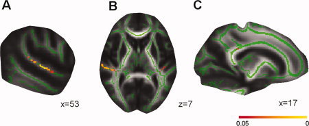
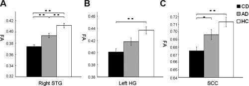
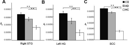
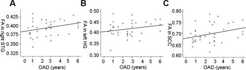
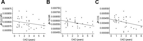
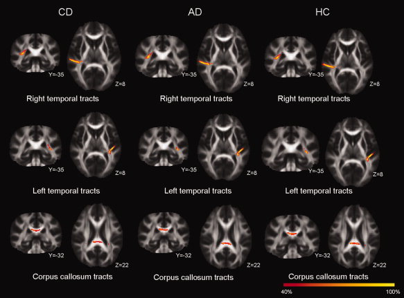
Previous studies have shown that white matter in the deaf brain changes due to hearing loss. However, how white-matter development is influenced by early hearing experience of deaf people is still unknown. Using diffusion tensor imaging and tract-based spatial statistics, we compared white-matter structures among three groups of subjects including 60 congenitally deaf individuals, 36 acquired deaf (AD) individuals, and 38 sex- and age-matched hearing controls (HC). The result showed that the deaf individuals had significantly reduced fractional anisotropy (FA) values in bilateral superior temporal cortex and the splenium of corpus callosum compared to HC. The reduction of FA values in acquired deafness correlated with onset age of deafness, but not the duration of deafness. To explore the underlying mechanism of FA changes in the deaf groups, we further analyzed radial and axial diffusivities and found that (1) the reduced FA values in deaf individuals compared to HC is primarily driven by higher radial diffusivity values and (2) in the AD, higher radial diffusivity was correlated with earlier onset age of deafness, but not the duration of deafness. These findings imply that early sensory experience is critical for the growth of fiber myelination, and anatomical reorganization following auditory deprivation is sensitive to early plasticity in the brain.


Figures

Figure 1
Figure 1
Differences in fractional anisotropy (FA) between congentially deaf (CD), acquired deaf (AD), and hearing controls (HC). The white‐matter skeleton (green) is projected onto the MNI brain (gray). The location of differences in FA value between CD, AD, and HC is indicated (Red–Yellow). (a) Areas in right superior temporal gyrus (STG) with difference in FA between CD, AD, and HC. (b) Areas in right STG (left side) and left Heschl's gyrus (right side) with differences in FA between CD, AD, and HC. The left side of the image corresponds to the participant's right hemisphere. (c) Areas in splenium of corpus callosum with differences in FA between CD, AD, and HC. A threshold‐free cluster enhancement corrected threshold of P < 0.05 is used. The color bar represents the P value.
Figure 2
Figure 2
Reduced fractional anisotropy (FA) in deaf individuals compared to hearing controls (HC). (a) Lower FA values in congentially deaf (CD) compared to acquired deaf (AD) and in both deaf groups compared to HC in right superior temporal gyrus (STG). (b) Lower FA values in CD compared to HC in left Heschl's gyrus (HG). (c) Lower FA values in CD compared to HC in splenium of corpus callosum (SCC). Values are presented in mean millimeters (standard error). *P < 0.05; **P < 0.01.
Figure 3
Figure 3
Increased radial diffusivity (λ) in deaf individuals compared to hearing controls. (a) Higher λ values in congenitally deaf (CD) compared to hearing controls (HC) as well as higher λ values in acquired deaf (AD) compared to HC in right superior temporal gyrus (STG). (b) Higher λ values in CD compared to HC as well as higher λ values in AD when compared with HC in left Heschl's gyrus (HG). (c) Higher λ values in CD and AD compared to HC in splenium of corpus callosum (SCC). Values are presented in mean millimeters (standard error). *p < 0.05; **p < 0.01.
Figure 4
Figure 4
Correlations between age of onset of deafness (AOD) in acquired deaf individuals and the mean fractional anisotropy (FA) in regions showing group FA differences. (a) Later AOD is correlated with higher FA values in right superior temporal gyrus (STG) (r = 0.286, P = 0.045). (b) Later AOD is correlated (not significantly) with higher FA values in left Heschl's gyrus (HG) (r = 0.233, P = 0.086). (c) Later, AOD is correlated with higher FA values in splenium of corpus callosum (SCC) (r = 0.306, P = 0.035). The correlations for right STG and left HG were still significant after partialing for duration of deafness (right STG: r = 0.318, P = 0.032; left HG: r = 0.324, P = 0.029).
Figure 5
Figure 5
Correlations between age of onset of deafness (AOD) in acquired deaf individuals and mean radial diffusivity (λ) values in regions showing group FA differences. (a) Later, AOD is correlated (not significantly) with lower λ values in right superior temporal gyrus (STG) (r = −0.235, P = 0.084). (b) Later, AOD is correlated with lower λ values in left Heschl's gyrus (HG) (r = 0.382, P = 0.011). (c) Later, AOD is correlated with lower λ values in splenium of corpus callosum (SCC) (r = 0.351, P = 0.018). The correlations for all three clusters were significant after partialing for duration of deafness (right STG: r = −0.352, P = 0.019; left HG: r = −0.432, P = 0.005; SCC: r = −0.295, P = 0.043).
Figure 6
Figure 6
Tractography distributions in congentially deaf (CD), acquired deaf (AD), and hearing controls (HC). The results were very similar for these three groups for right superior temporal gyrus (STG, top row), left Heschl's gyrus (HG, middle row), and splenium of corpus callosum (SCC, bottom row). The color bar represents the percent of participants having positive results from fiber tracking for that voxel.

References

    1. Allen JS, Emmorey K, Bruss J, Damasio H ( 2008): Morphology of the insula in relation to hearing status and sign language experience. J Neurosci 28: 11900–11905. - PMC - PubMed
    1. Auer ET Jr, Bernstein LE, Sungkarat W, Singh M ( 2007): Vibrotactile activation of the auditory cortices in deaf versus hearing adults. Neuroreport 18: 645–648. - PMC - PubMed
    1. Barnea‐Goraly N, Menon V, Eckert M, Tamm L, Bammer R, Karchemskiy A, Dant CC, Reiss AL ( 2005): White matter development during childhood and adolescence: A cross‐sectional diffusion tensor imaging study. Cereb Cortex 15: 1848–1854. - PubMed
    1. Basser PJ, Pierpaoli C ( 1996): Microstructural and physiological features of tissues elucidated by quantitative‐diffusion‐tensor MRI. J Magn Reson B 111: 209–219. - PubMed
    1. Basser PJ, Mattiello J, LeBihan D ( 1994): Estimation of the effective self‐diffusion tensor from the NMR spin echo. J Magn Reson B 103: 247–254. - PubMed

Publication types