. 2011 Mar;6(2):171-8.
doi: 10.2217/rme.10.103.

Cardiac tumorigenic potential of induced pluripotent stem cells in an immunocompetent host with myocardial infarction

Affiliations

Cardiac tumorigenic potential of induced pluripotent stem cells in an immunocompetent host with myocardial infarction

Rafeeq P H Ahmed et al. Regen Med. 2011 Mar.

Abstract

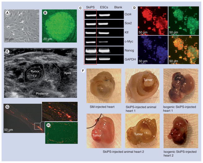
Aim: Genetic reprogramming of somatic cells with stemness genes to restore their pluripotent status is being studied extensively to generate pluripotent stem cells as an alternative to embryonic stem cells. This study was designed to examine the effectiveness of skeletal myoblast-derived induced pluripotent stem cells (SkiPS) from young male Oct4/GFP transgenic mice for regeneration of the infarcted heart.

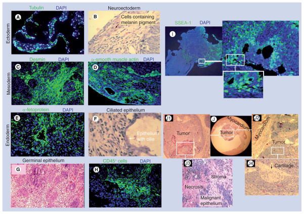
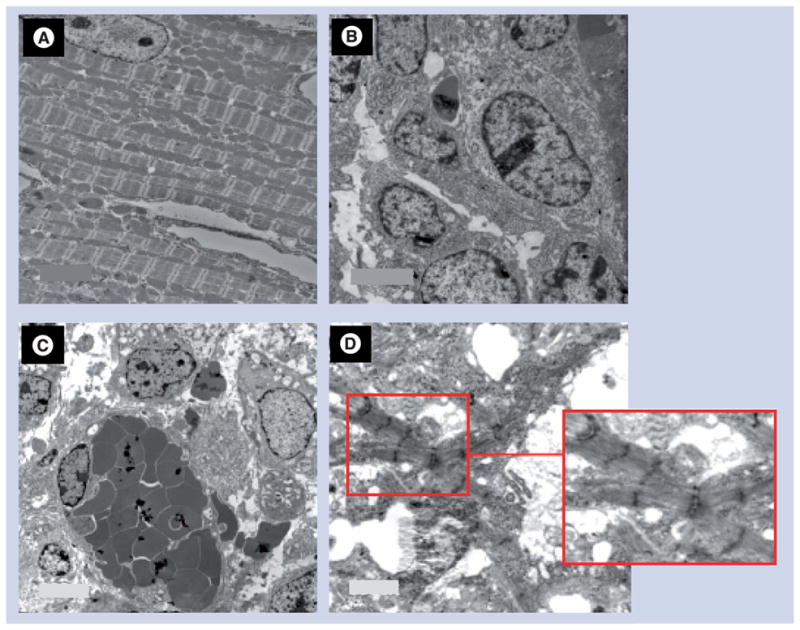
Methods & results: A mouse model of permanent coronary artery ligation was developed in young female immunocompetent C57BL/6J or C57BL/6x129S4 SV/jae Oct4/GFP mice. SkiPS labeled with Q-dots (3 × 10(5) in 10 µl basal Dulbecco's modified Eagle's medium) were transplanted in and around the area of infarct immediately after coronary artery ligation (n = 16) under direct vision. Control mice (n = 12) were injected with the same number of skeletal myoblasts. Histological studies documented successful engraftment of SkiPS in all the surviving animals 4 weeks later. However, six of the 16 SkiPS-transplanted (37.5%) animal hearts showed intramural teratomas, whereas no tumor growth was observed in the control mice. Q-dot-labeled donor cells were also observed at the site of tumors. Histological studies revealed that teratomas were composed of cells from all of the three embryonic germ layers. Ultra-structure studies confirmed the histological findings and showed regions with well-organized myofibrillar structures in the tumors.

Conclusion: Undifferentiated induced pluripotent stem cells should not be recommended for cardiac transplantation unless screened for specific teratogenic precursors or predifferentiated into cardiac lineage prior to transplantation.


Figures

Figure 1
Figure 1. Reprogramming of mouse skeletal myoblasts
(A) Primary culture of SMs isolated from a male donor Oct4/GFP transgenic mouse and (B) mouse SkiPS with typical ESC-like morphology on day 20 after retroviral transduction of Yamanaka’s quartet of stemness factors. (C) Reverse transcriptase-PCR showing endogenous expression of stemness factors in SkiPS, which was similar to mouse ESCs. (D) SkiPS identified on the basis of Oct4/GFP expression (green) immunostained positively for stage-specific embryonic antigen-1 (red fluorescence) to show their pluripotent status. Nuclei were visualized by staining with 4′-6-diamidino-2-phenylindole (original magnification = 400×). (E) Transthoracic ECG demonstrating cardiac tumor in isogenic SkiPS-treated animal heart at 4 weeks after treatment. (F) Macroscopic view of the mouse hearts showing teratoma formation at 4 weeks after allogenic and isogenic SkiPS engraftment, whereas no teratoma was observed in SM-transplanted animal hearts. (G & H) Photomicrographs of the histological sections of mouse heart 1 showing extensive presence of Q-dot-labeled SkiPS (red fluorescence) in (G) infarct and (H) tumor areas. White box in (G) has been magnified to show the presence of Q-dot-labeled SkiPS. ESC: Embryonic stem cell; LV: Left ventricle; SkiPS: Skeletal myoblast-derived induced pluripotent stem cells; SM: Skeletal myoblasts.
Figure 2
Figure 2. Differentiation of skeletal myoblast-derived induced pluripotent stem cells into teratoma in the infarcted heart
Histological studies of skeletal myoblast-derived induced pluripotent stem cell-derived teratomas in the myocardium showing derivatives of ectoderm ((A) tubulin and (B) neuroectoderm), mesoderm ((C) desmin and (D) smooth muscle actin), and endoderm ((E) α-fetoprotein and (F) ciliated epithelium). (A & C–E) show immunostaining of the histological sections with specific antibody, whereas (B & F) show hematoxylin and eosin staining. (G) Extensive presence of duct-like structures lined with germinal epithelium were observed in tumor number 2 (original magnification = 400 ×). (H) Fluorescence immunostaining of histological sections showing infiltration of CD45-positive cells at 4 weeks after skeletal myoblast-derived induced pluripotent stem cell engraftment. (I) Fluorescence immunostaining for stage SSEA-1-expressing pluripotent cells in the tumor. (J) The microscopic view of tumor number 1 occupying the left ventricle cavity. White boxes in (J) have been magnified as (J1 & J2) to show a larger view of the teratoma and its association with the myocardium, respectively, whereas magnified regions in (J3 & J4) show necrosis and malignant epithelium and cartilage, respectively. DAPI: 4′,6-diamidino-2-phenylindole; SSEA: Stage-specific embryonic antigen.
Figure 3
Figure 3. Transmission electron microscopic studies 4 weeks after skeletal myoblast-derived induced pluripotent stem cell engraftment in a mouse heart
(A) Transmission electron micrograph of healthy myocardium in a skeletal myoblast-derived induced pluripotent stem cell-transplanted mouse heart showing a normal myocyte with orderly arranged sarcomeres and mitochondria. (B) Tumor cells in the center of the tumor. (C) Russell body development characterized by accumulation of proteinaceous material occupying most of the cytoplasm with typically marginated nucleus. (D) Although we observed relatively less organized myofibrillar bundles in some part of the tumors, a more mature sarcomeric organization was present in some regions. (Please see the red box, which depicts a magnified image of the selected region in (D) to highlight the sarcomeric organization in the selected region.) Original magnifications: (A) 6000×, (B) 15000×, (C) 8000× and (D) 8000×.

References

    1. Takahashi K, Yamanaka S. Induction of pluripotent stem cells from mouse embryonic and adult fibroblast cultures by defined factors. Cell. 2006;126:663–676. - PubMed
    1. Lowry WE, Plath K. The many ways to make an iPS cell. Nat Biotechnol. 2008;26:1246–1248. - PubMed
    1. Kuzmenkin A, Liang H, Xu G, et al. Functional characterization of cardiomyocytes derived from murine induced pluripotent stem cells in vitro. FASEB J. 2009;23:4168–4180. - PubMed
    1. Nelson TJ, Martinez-Fernandez A, Yamada S, et al. Repair of acute myocardial infarction by human stemness factors induced pluripotent stem cells. Circulation. 2009;120:408–416. - PMC - PubMed
    1. Niagara MI, Haider H, Jiang S, et al. Pharmacologically preconditioned skeletal myoblasts are resistant to oxidative stress and promote angiomyogenesis via release of paracrine factors in the infarcted heart. Circ Res. 2007;100:545–555. - PubMed

Publication types

MeSH terms

Substances