. 2011 Mar 11:8:113.
doi: 10.1186/1743-422X-8-113.

Tim-3 expression on peripheral T cell subsets correlates with disease progression in hepatitis B infection

Affiliations

Tim-3 expression on peripheral T cell subsets correlates with disease progression in hepatitis B infection

Wei Wu et al. Virol J. .

Abstract

Background and objective: T-cell immunoglobulin domain and mucin domain-containing molecule-3 (Tim-3) represents a novel mechanism of T-cell dysfunction in chronic viral diseases. However, the role of Tim-3 in the pathogenesis of chronic hepatitis B (CHB) is not well understood. We investigated Tim-3 expression on peripheral T cell subsets and analyzed the relationship between Tim-3 expression and disease progression in HBV infection.

Methods: peripheral blood samples were obtained from CHB patients (n = 40), including 23 patients with moderate CHB [MCHB] and 17 with severe CHB [SCHB]. Control samples were obtained from nine acute hepatitis B patients (AHB) and 26 age-matched healthy subjects. The expression of Tim-3 on T cells was determined by flow cytometry.

Results: Tim-3 expression was elevated on peripheral CD4+ and CD8+ T cells from AHB and CHB patients compared to those from healthy controls. The percentage of Tim-3+ T cells was further increased in SCHB patients relative to MCHB patients and showed a positive correlation with conventional markers for liver injury (alanine aminotransferase (ALT), aspartate transaminase (AST), total bilirubin (TB) and international normalized ratio (INR) level). The frequency of Tim-3-expressing T cells was negatively correlated with T-bet mRNA expression and plasma interferon-gamma (INF-gamma) levels. Further, Tim-3 expression on CD4+ or CD8+ T cells was reduced in CHB patients with disease remission after antiviral treatment and in AHB patients during the convalescence phase.

Conclusions: Our results suggest that over-expression of Tim-3 is involved in disease progression of CHB and that Tim-3 may participate in skewing of Th1/Tc1 response, which contributes to persistency of HBV infection.


Figures

Figure 1
Figure 1
Elevated Tim-3 expression on circulating CD4 and CD8 T cells in HBV-infected individuals. (A) PBMCs from normal controls, AHB patients (early phase), MCHB and SCHB patients were stained with antibodies against CD3, CD4/CD8, and Tim-3. An isotype-matched antibody was used as a negative control. (B) The percentage of Tim-3 cells within CD4+ and CD8+ T cell populations are increased in AHB and CHB patients and further enhanced in the severe stage of CHB. Each dot represents an individual data point and the horizontal lines represent the mean. The Mann-Whitney U test was used to compare differences among groups. (C) There is a significantly positive correlation between the frequency of Tim-3-expressing CD4+ and CD8+ T cells in the studied subjects. Spearman test was performed for correlation analysis.
Figure 2
Figure 2
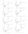
Association between the frequency of Tim-3+ T cells and conventional markers for liver damage in CHB patients. The frequency of CD4+ or CD8+ T cells expressing Tim-3 are positively correlated with the levels of laboratory parameters for assessing liver injury such as ALT (A), AST (B), TB (C) and INR (D). Spearman test was used for correlation analysis.
Figure 3
Figure 3
Association between the frequency of Tim-3+ T cells and T-bet mRNA expression in CHB patients. (A) T-bet mRNA expression was determined in patients separated into four groups: normal controls (N = 12), AHB patients (early phase) (N = 7), MCHB patients (N = 15) and SCHB patients (N = 10). The bars represent the (95% CI) T-bet mRNA expression for each group and the differences among groups were analyzed using the Mann-Whitney U test. (B) Spearman correlation analysis showed a negative association between the frequency of Tim-3-postivie T cells and T-bet mRNA expression.
Figure 4
Figure 4
Association between the frequency of Tim-3+ T cells and plasma IFN-gamma level in CHB patients. (A) IFN-gamma was measured in plasma from normal controls (N = 12), AHB patients (early phase) (N = 7), MCHB patients (N = 15) and SCHB patients (N = 10). The bars represent the (95% CI) IFN-gamma level for each group and the differences among groups were analyzed using the Mann-Whitney U test. (B) Spearman correlation analysis showed there were negative associations between the frequency of Tim-3-postivie T cells and plasma IFN-gamma level.
Figure 5
Figure 5
longitudinal analysis of Tim-3 expression in CHB and AHB patients. (A) The percentage of Tim-3+ T cells decreased in CHB patients after successful antiviral treatment. The frequency of Tim-3+ cells in CD4+ and CD8+ T cells before and after treatment is shown. (B) The frequency of Tim-3+ T cells decreased in AHB patients during disease convalescence. The frequency of Tim-3+ cells in CD4+ and CD8+ T cells at early stage and convalescence stage is shown.

References

    1. Lok ASF, McMahon BJ. Chronic hepatitis B: Update 2009. Hepatology. 2009;50:661–662. doi: 10.1002/hep.23190. - DOI - PubMed
    1. Rehermann B, Fowler P, Sidney J, Person J, Redeker A, Brown M, Moss B, Sette A, Chisari FV. The cytotoxic T lymphocyte response to multiple hepatitis B virus polymerase epitopes during and after acute viral hepatitis. J Exp Med. 1995;181:1047–58. doi: 10.1084/jem.181.3.1047. - DOI - PMC - PubMed
    1. Chisari FV, Isogawa M, Wieland SF. Pathogenesis of hepatitis B virus infection. Pathol Biol (Paris) 2010;58:258–66. - PMC - PubMed
    1. Barber DL, Wherry EJ, Masopust D, Zhu B, Allison JP, Sharpe AH, Freeman GJ, Ahmed R. Restoring function in exhausted CD8 T cells during chronic viral infection. Nature. 2006;439:682–7. doi: 10.1038/nature04444. - DOI - PubMed
    1. Day CL, Kaufmann DE, Kiepiela P, Brown JA, Moodley ES, Reddy S, Mackey EW, Miller JD, Leslie AJ, DePierres C, Mncube Z, Duraiswamy J, Zhu B, Eichbaum Q, Altfeld M, Wherry EJ, Coovadia HM, Goulder PJ, Klenerman P, Ahmed R, Freeman GJ, Walker BD. PD-1 expression on HIV-specific T cells is associated with T-cell exhaustion and disease progression. Nature. 2006;443:350–4. doi: 10.1038/nature05115. - DOI - PubMed

Publication types

MeSH terms