. 2011 Mar;17(3):449-56.
doi: 10.3201/eid1703.100526.

Amplification of emerging viruses in a bat colony

Affiliations

Amplification of emerging viruses in a bat colony

Jan Felix Drexler et al. Emerg Infect Dis. 2011 Mar.

Abstract

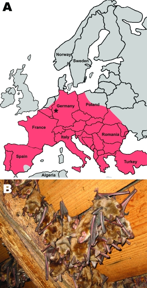
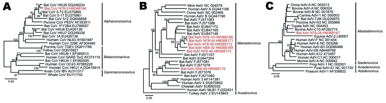
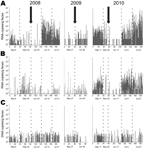
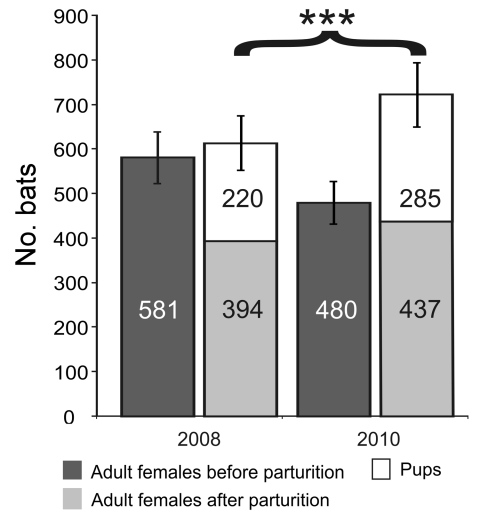
Bats host noteworthy viral pathogens, including coronaviruses, astroviruses, and adenoviruses. Knowledge on the ecology of reservoir-borne viruses is critical for preventive approaches against zoonotic epidemics. We studied a maternity colony of Myotis myotis bats in the attic of a private house in a suburban neighborhood in Rhineland-Palatinate, Germany, during 2008, 2009, and 2010. One coronavirus, 6 astroviruses, and 1 novel adenovirus were identified and monitored quantitatively. Strong and specific amplification of RNA viruses, but not of DNA viruses, occurred during colony formation and after parturition. The breeding success of the colony was significantly better in 2010 than in 2008, in spite of stronger amplification of coronaviruses and astroviruses in 2010, suggesting that these viruses had little pathogenic influence on bats. However, the general correlation of virus and bat population dynamics suggests that bats control infections similar to other mammals and that they may well experience epidemics of viruses under certain circumstances.


Figures

Figure 1
Figure 1
A) Location of studied maternity bat roost (indicated by asterisk) in the state of Rhineland-Palatinate, Germany (50°25′46.91′′N, 6°55′52.17′′E). Red shading indicates the distribution of the studied bat species (adapted from the IUCN Red List of Threatened Species, v. 2010; www.iucnredlist.org). B) Cluster of Myotis myotis female bats hanging from the roof interior.
Figure 2
Figure 2
Phylogenetic relationships of novel bat viruses. A) Coronavirus, B) astrovirus, C) adenovirus. Neighbor-joining phylogenies were generated with MEGA (www.megasoftware.net), by using an amino acid percentage distance substitution model drawn to scale, complete deletion option, and 1,000 bootstrap reiterations. Bootstrap values are shown next to the branches; values <65 were removed for graphic reasons. Viruses newly identified in this study are shown in red. Viral genera are depicted next to taxon names. The BLOSUM aligned datasets corresponded to an 816-nt alignment, corresponding to nucleotides 14,781–15,596 in severe acute respiratory syndrome coronavirus (SARS-CoV) strain Tor2 (GenBank accession no. AY274119) for CoVs (A); a 381-nt alignment corresponding to nt 3,437–3,817 in mink astrovirus (AstV) (GenBank NC_004579) for AstVs (B); and to a 255-nt alignment corresponding to nt 46–300 in the bovine adenovirus (AdV) C10 hexon gene (GenBank accession no. AF282774) for AdVs (C). Trees were visualized in MEGA4, with prototype virus sequences restricted to ≈20 taxa additional to newly identified viruses for graphic reasons. Scale bars indicate amino acid substitutions per site.
Figure 3
Figure 3
Detection frequency of bat viruses and virus nucleic acid concentrations over time. A) Coronavirus; B) astrovirus; C) adenovirus. Samples were obtained approximately every 3 weeks from the same Myotis myotis bat maternity roost in 3 different sampling years, 2008–2010. Each sample was tested by specific real-time reverse transcription–PCR (RT-PCR) with RNA/DNA concentrations per gram of feces given on the y axis. The arrows indicate the time of birth of the first pup. Numbers on the x-axis represent individual fecal pools tested, consisting of 5 single fecal pellets each. Five different sampling dates (below each panel) are shown by dotted lines for each sampling year. Empty columns indicate pools that tested negative. In panel B, light and dark gray bars identify results by 2 different real-time RT-PCRs that were used simultaneously to cover the large astrovirus diversity encountered.
Figure 4
Figure 4
Myotis myotis bat maternity roost composition and reproductive success. Age composition of bats composing the M. myotis maternity roost under study are depicted before and after parturition in 2 different sampling years, 2008 and 2010. The y-axis represents the number of individual bats, additionally indicated in individual bars. The brace and asterisks represent statistical significance of the gain in total colony size after parturition in 2010, compared with colony size in 2008. Error bars represent an assumed 10% error margin in counting.

References

    1. Simmons NB. Order Chiroptera. In: Wilson DE, Reeder DM, editors. Mammal species of the world: a taxonomic and geographic reference. Baltimore: Johns Hopkins University Press; 2005. p. 312–529.
    1. Calisher CH, Childs JE, Field HE, Holmes KV, Schountz T. Bats: important reservoir hosts of emerging viruses. Clin Microbiol Rev. 2006;19:531–45.10.1128/CMR.00017-06 - DOI - PMC - PubMed
    1. Dobson AP. Virology. What links bats to emerging infectious diseases? Science. 2005;310:628–9.10.1126/science.1120872 - DOI - PubMed
    1. Chu DK, Peiris JS, Chen H, Guan Y, Poon LL. Genomic characterizations of bat coronaviruses (1A, 1B and HKU8) and evidence for co-infections in Miniopterus bats. J Gen Virol. 2008;89:1282–7.10.1099/vir.0.83605-0 - DOI - PubMed
    1. Wibbelt G, Kurth A, Yasmum N, Bannert M, Nagel S, Nitsche A, et al. Discovery of herpesviruses in bats. J Gen Virol. 2007;88:2651–5.10.1099/vir.0.83045-0 - DOI - PubMed

Publication types

MeSH terms

LinkOut - more resources