. 2011 Mar;17(3):480-7.
doi: 10.3201/eid1703.101410.

Novel picornavirus in Turkey poults with hepatitis, California, USA

Affiliations

Novel picornavirus in Turkey poults with hepatitis, California, USA

Kirsi S Honkavuori et al. Emerg Infect Dis. 2011 Mar.

Abstract

To identify a candidate etiologic agent for turkey viral hepatitis, we analyzed samples from diseased turkey poults from 8 commercial flocks in California, USA, that were collected during 2008-2010. High-throughput pyrosequencing of RNA from livers of poults with turkey viral hepatitis (TVH) revealed picornavirus sequences. Subsequent cloning of the ≈9-kb genome showed an organization similar to that of picornaviruses with conservation of motifs within the P1, P2, and P3 genome regions, but also unique features, including a 1.2-kb sequence of unknown function at the junction of P1 and P2 regions. Real-time PCR confirmed viral RNA in liver, bile, intestine, serum, and cloacal swab specimens from diseased poults. Analysis of liver by in situ hybridization with viral probes and immunohistochemical testing of serum demonstrated viral nucleic acid and protein in livers of diseased poults. Molecular, anatomic, and immunologic evidence suggests that TVH is caused by a novel picornavirus, tentatively named turkey hepatitis virus.


Figures

Figure 1
Figure 1
Predicted turkey hepatitis virus (THV) genome organization based on sequence comparison to known picornaviruses. Dotted lines above the genome depict the location of the original sequences obtained by high-throughput sequence analysis. Conserved picornaviral motifs and predicted potential cleavage sites along the coding region are indicated in the bar below.
Figure 2
Figure 2
Relationships between turkey hepatitis virus (THV) and other picornaviruses. The phylogenetic analyses were based on amino acid sequences of the combined 2C, 3C, and 3D regions (A), the P1 region (B), and complete coding regions, excluding divergent aa 799–1199 (C). Representative sequences from different picornavirus genera and recently discovered, unclassified viruses were obtained from GenBank; accession numbers are indicated. Bootstrap values are given at the respective nodes; scale bars indicate number of amino acid substitutions per site. TMEV, Theiler’s murine encephalomyelitis virus; EMCV, encephalomyocarditis virus; SVV, Seneca Valley virus; HCoSV-A1, human cosavirus A1; HCoSV-D1, human cosavirus D1; ERBV, equine rhinitis B virus; ERAV, equine rhinitis A virus; BRBV, bovine rhinitis B virus; FMDV, foot-and-mouth disease virus; PTV, porcine teschovirus; PSV, porcine sapelovirus; SSV, simian sapelovirus; ASV, avian sapelovirus; HRV-C, human rhinovirus C; CV-A21, coxsackievirus A21; EV-96, enterovirus 96; TV-2, turdivirus 2; TV-3, turdivirus 3; TV-1, turdivirus 1; AiV, Aichi virus; BKV, bovine kobuvirus; PKV, porcine kobuvirus; AEV, avian encephalomyelitis virus; HAV, hepatitis A virus; SePV, seal picornavirus; DHAV, duck hepatitis A virus; HPeV, human parechovirus; LV, Ljungan virus.
Figure 3
Figure 3
Results of in situ hybridization experiments. Hybridization of β-actin–specific and turkey hepatitis virus (THV)–specific oglionucleotide probes with FastRed staining on hepatitis-affected liver tissue from poult 2993A (A and B, respectively) and on nondiseased liver tissue from poult 1927B (C and D, respectively). Brightfield microscopy images; original magnification ×40.
Figure 4
Figure 4
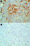
Immunohistologic staining of liver tissues with serum from a turkey poult with turkey viral hepatitis (TVH). Serum sample from PCR-positive poult 394.9 demonstrates turkey hepatitis virus (THV) antigens in clusters of cells in liver tissue of TVH-affected poult 2993A (A) but not in liver sections from nondiseased poult 1927B (B). Sections were counterstained with hematoxylin. Brightfield microscopy images; original magnification ×40.

References

    1. Klein PN, Castro AE, Meteyer CU, Reynolds B, Swartzman-Andert JA, Cooper G, et al. Experimental transmission of turkey viral hepatitis to day-old poults and identification of associated viral particles resembling picornaviruses. Avian Dis. 1991;35:115–25. 10.2307/1591303 - DOI - PubMed
    1. Snoeyenbos GH, Basch HI. Further studies of virus hepatitis of turkeys. Avian Dis. 1960;3:477–84. 10.2307/1587700 - DOI
    1. Mongeau JD, Truscott RB, Ferguson AE, Connell MC. Virus hepatitis in turkeys. Avian Dis. 1959;3:388–96. 10.2307/1587578 - DOI
    1. Snoeyenbos GH, Basch HI, Sevoian M. An infectious agent producing hepatitis in turkeys. Avian Dis. 1959;3:377–88. 10.2307/1587577 - DOI
    1. Tzianabos T, Snoeyenbos GH. Some physiochemical properties of turkey hepatitis virus. Avian Dis. 1965;9:152–6. 10.2307/1587802 - DOI - PubMed

Publication types

LinkOut - more resources