. 2011 May 5;117(18):4773-7.
doi: 10.1182/blood-2011-01-330514. Epub 2011 Mar 10.

Hematopoietic stem/progenitor cells, generation of induced pluripotent stem cells, and isolation of endothelial progenitors from 21- to 23.5-year cryopreserved cord blood

Affiliations

Hematopoietic stem/progenitor cells, generation of induced pluripotent stem cells, and isolation of endothelial progenitors from 21- to 23.5-year cryopreserved cord blood

Hal E Broxmeyer et al. Blood. .

Abstract

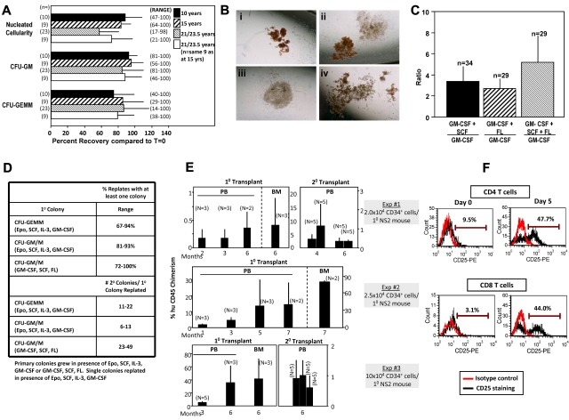
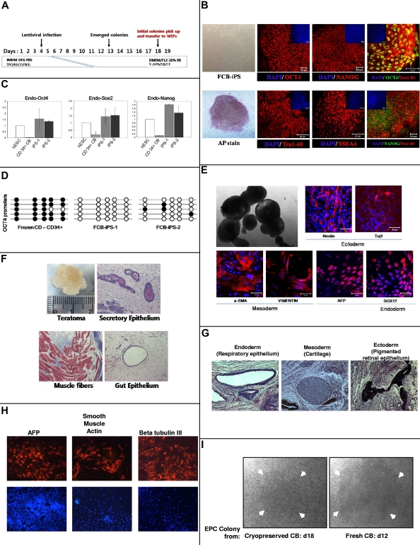
Cryopreservation of hematopoietic stem cells (HSCs) and hematopoietic progenitor cells (HPCs) is crucial for cord blood (CB) banking and transplantation. We evaluated recovery of functional HPC cryopreserved as mononuclear or unseparated cells for up to 23.5 years compared with prefreeze values of the same CB units. Highly efficient recovery (80%-100%) was apparent for granulocyte-macrophage and multipotential hematopoietic progenitors, although some collections had reproducible low recovery. Proliferative potential, response to multiple cytokines, and replating of HPC colonies was extensive. CD34(+) cells isolated from CB cryopreserved for up to 21 years had long-term (≥ 6 month) engrafting capability in primary and secondary immunodeficient mice reflecting recovery of long-term repopulating, self-renewing HSCs. We recovered functionally responsive CD4(+) and CD8(+) T lymphocytes, generated induced pluripotent stem (iPS) cells with differentiation representing all 3 germ cell lineages in vitro and in vivo, and detected high proliferative endothelial colony forming cells, results of relevance to CB biology and banking.


Figures

Figure 1
Figure 1
Recovery of nucleated cellularity, HPCs, HSCs, and immune cells after cryopreservation and long-term frozen storage of CB. (A) Comparative percent recovery of nucleated cells, CFU-GM, and CFU-GEMM compared with prefreeze numbers for 10, 15, and 21-23.5 years of the exact sample frozen. n = number of different samples thawed for analysis. Results shown as mean ± 1 SEM with range of recoveries shown in parentheses. (B) Representative examples of colonies grown from CB thawed after 21-23.5 years in frozen state: (i) CFU-GEMM colony; (ii) CFU-GEMM (left) and CFU-GM (right) colonies; (iii) CFU-GM colony; (iv) CFU-GEMM colony. A Nikon TMS microscope was used with a PLAN objective 4× and projection lens magnification of 2.5× for a total magnification of 10× at 25°C. We used 35mm film (ASA200), and pictures were made with a Nikon HFX-DX automatic 35mm camera system. (C) Ratio of CFU-GM colonies formed after stimulation of CB cells with GM-CSF plus either SCF, FL, or SCF plus FL, divided by number of colonies formed by stimulation of same cells with only GM-CSF. n = number of different CB samples analyzed from cells frozen from 21-23.5 years before thaw and analysis. (D) Replating capacity of single CFU-GEMM or CFU-GM plus CFU-M (macrophage) colonies recovered from thawed CB cells stored frozen for up to 21 years. Results shown are from a total of over 1000 separately replated colonies each, and designated percent of secondary plates with at least 1 colony (% replates = top) and range of colonies in secondary dish per single replated colony (= bottom). (E) Analysis of engrafting capacity in sublethally irradiated NOD-SCID IL-2 receptor γ chain null (NSG) mice of cells (≥ 90% CD34+) purified from unseparated CB stored frozen for 18-21 years before thawing. Each experiment shows chimerism data from a different frozen CB unit. Analysis of human CD45+chimerism in peripheral blood (PB) or bone marrow (CB) of primary (1°) mouse recipients with N = number of mice per group, or CD45+ cell chimerism in secondary (2°) recipients of the same mouse strain given the same sublethal irradiation dose. For 2° recipients, each bar represents the number of secondary recipients per pooled BM of 1° recipient mice (experiments 1 and 2 only). Results are given as mean ± 1 SEM. (F) Response of CD4+ and CD8+ T lymphocytes, (≥ 98% Pure) isolated from thawed unseparated CB cells stored frozen for 21 years, as assessed by flow cytometry, to anti-CD3/CD28 stimulation, as determined by induced expression of CD25, a T-cell activation antigen. Shown are experiments from 1 of 2 experiments in which different frozen CB units were assessed.
Figure 2
Figure 2
Reprogramming of 21-year-old frozen human CB CD34+ cells to iPS cells (A-H) and recovery of ECFCs (I). The phase contrast images in panels B (FCB-iPS and AP stain), E (top left image), and I were viewed using a Zeiss Axiovert 25 CFL inverted microscope with a 5× CP-ACHROMAT/0.12 NA objective. Images were acquired using a SPOT RT color camera (Diagnostic Instruments) with the manufacturer's software. Phase contrast images were taken with air objectives. The confocal images in panels B and E were viewed with a Zeiss Axiovert 100 LSM 510 confocal microscope using as objective a C-Apochromat (at 40×/1.2W corr) or Plan-Apochromat (at 10×/0.45). Images were taken by epifluorescence detector and transillumination detector and processed using Zeiss LSM Image Browser Version 3.5.0.376 software. All the images were taken at room temperature. Cells were mounted in proLong Gold antifade mounting reagent with DAPI (Invitrogen) and confocal images were taken using water immersion objectives. (A) Schematic representation of strategy used at IU to reprogram CD34+ cells from frozen human CB. TPO indicates Thrombopoietin; hSCF, stem cell factor; Flt2L, fms-like tyrosine kinase 3 ligand; Y, Y-27632; VPA, valporic acid; and SB, SB202190. (B) Immunocytochemistry for pluripotency markers OCT4 (Alexa 448 or 546 nm), NANOG (Alexa 488 or 564 nm), TRA-1–60 (Alexa 564 nm), SSEA4 (Alexa564 nm) and alkaline phosphatase (AP). Small panels on top part of subpanels show total cell content per field stained with DAPI. Scale bars represent 50 μm. (C) Quantitative RT-PCR analysis for expression of endogenous ES cell-marker genes OCT4, SOX2, and NANOG in hESC line H9, 2 iPS cell lines generated, and parental CD34+ cells. Specific primers were designed to probe for 3′ untranslated region (Endo) to measure expression of the endogenous gene. Individual PCR reactions were normalized against β-actin and plotted relative to expression levels in hESCs. (D) Each horizontal row of circles represents an individual sequencing reaction for a given amplicon. Open and filled circles, respectively, represent unmethylated and methylated CpGs dinucleotides. (E) Morphology of 10-day embryoid body (EB) under phase contrast microscopy (top left). Immunostaining of frozen CB (FCB)–iPS derivatives on day 10 of differentiation revealed expression of ectodermal (Nestin and Tuj1), mesodermal (α-SMA and Vimentin), and endodermal (AFP and Sox17) marker proteins (all with Alexa 546nm). Nuclei are stained with DAPI (blue). Scale bars represent 50 μm. (F) Teratomas derived from immunodeficient mice injected with FCB-iPS cells shows tissues representing all 3 embryonic germ layers, including secretory epithelium (ectoderm), muscle fibers (mesoderm), and gut epithelium (endoderm). Samples from the teratomas were paraffin-embedded and serially sectioned (5 μm) using a microtome (Leica Microsystems). To analyze the 3 germ-layer lineage cells derived from injected FCB-iPS cells in the teratomas, sectioned slides were histologically examined by hematoxylin and eosin for the gut epithelium and special stains as follows: PAS stain for the secretory epithelium, and Masson trichrome stain for muscle fibers. Images were taken with a Nikon Eclipse 50i equipped with a digital camera (Infinty 2 Megapixel; Lumenera Corp) at a magnification of Plan Fluor 10×/0.30 and analyzed using the i-solution image analysis program (iMT; i-Solution Inc). Teratoma images were taken with air objectives. Results in panels B through F were from studies performed at IU. Results in panels G and H were from another 21-year cryopreserved CB sample sent to, thawed, and separately reprogrammed at Johns Hopkins Medical Center after isolation of CD34+ cells. For studies in panels G and H, a Nikon Eclipse TE2000-U microscope was used, with ELWD Plan Fluor, NA:0.45 at 25°C. For panel G, fixed slides were used with H&E staining, and for panel H, PBS plus 0.1% BSA was used with Alexa 555 and DAPI. Images were taken with Imaging Micropublisher 5.0 (Q Imaging) camera with Q Capture Version 3.1.2 (Q Imaging) software. Markers for teratoma formation in vivo (G) and for differentiation of EBs from reprogrammed iPS cells in vitro (H). (I) Representative ECFC colony from MNCs isolated from 21-year CB defrosts and cultured for 18 days (left) compared with colony from freshly obtained CB cultured for 12 days. Arrows denote size of colonies. Note that even after 18 days culture of frozen CB-derived colonies, the colony size is not as large as that of colony from fresh CB at 12 days of culture.

References

    1. Gluckman E, Broxmeyer HA, Auerbach AD, et al. Hematopoietic reconstitution in a patient with Fanconi's anemia by means of umbilical-cord blood from an HLA-identical sibling. N Engl J Med. 1989;321(17):1174–1178. - PubMed
    1. Broxmeyer HE, Douglas GW, Hangoc G, et al. Human umbilical cord blood as a potential source of transplantable hematopoietic stem/progenitor cells. Proc Natl Acad Sci U S A. 1989;86(10):3828–3832. - PMC - PubMed
    1. Rocha V, Broxmeyer HE. New approaches for improving engraftment after cord blood transplantation. Biol Blood Marrow Transplant. 2010;16:S126–S132. - PubMed
    1. Wagner JE, Barker JN, DeFor TE, et al. Transplantation of unrelated donor umbilical cord blood in 102 patients with malignant and nonmalignant diseases: influence of CD34 cell dose and HLA disparity on treatment-related mortality and survival. Blood. 2002;100(5):1611–1618. - PubMed
    1. Kurtzberg J, Prasad VK, Carter SL, et al. Results of the Cord Blood Transplantation Study (COBLT): clinical outcomes of unrelated donor umbilical cord blood transplantation in pediatric patients with hematologic malignancies. Blood. 2008;112(10):4318–4327. - PMC - PubMed

Publication types

MeSH terms

Substances