. 2011 Mar 11;331(6022):1336-9.
doi: 10.1126/science.1199633.

A polarized epithelium organized by beta- and alpha-catenin predates cadherin and metazoan origins

Affiliations

A polarized epithelium organized by beta- and alpha-catenin predates cadherin and metazoan origins

Daniel J Dickinson et al. Science. .

Abstract

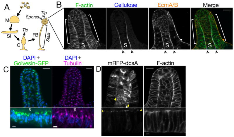
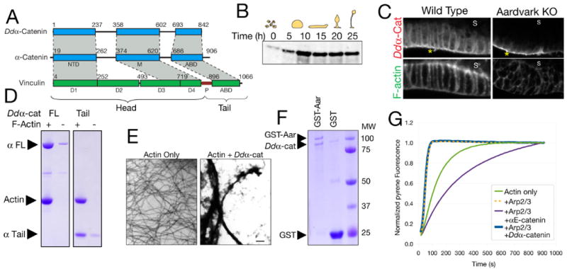
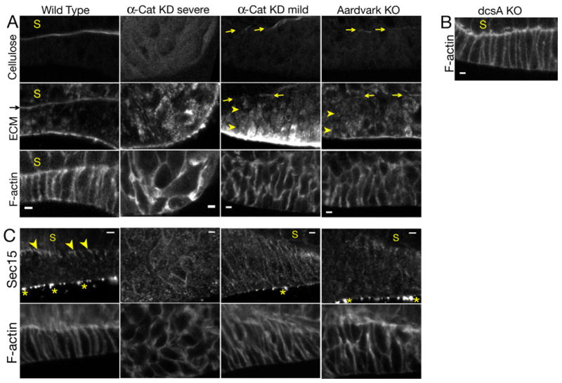
A fundamental characteristic of metazoans is the formation of a simple, polarized epithelium. In higher animals, the structural integrity and functional polarization of simple epithelia require a cell-cell adhesion complex that contains a classical cadherin, the Wnt-signaling protein β-catenin and the actin-binding protein α-catenin. We show that the non-metazoan Dictyostelium discoideum forms a polarized epithelium that is essential for multicellular development. Although D. discoideum lacks a cadherin homolog, we identify an α-catenin ortholog that binds a β-catenin-related protein. Both proteins are essential for formation of the epithelium, polarized protein secretion, and proper multicellular morphogenesis. Thus, the organizational principles of metazoan multicellularity may be more ancient than previously recognized, and the role of the catenins in cell polarity predates the evolution of Wnt signaling and classical cadherins.


Figures

Figure 1
Figure 1
A) D. discoideum developmental process. Abbreviations: M, mound; Sl, slug; C, culminant; FB, fruiting body. B) Confocal section of the tip of a wild-type culminant. Brackets indicate the tip epithelium; arrowheads indicate the stalk tube; S indicates the stalk. C) Maximum intensity projections showing Golgi (left), centrosomes (right) and nuclei (DAPI stain) in the entire tip (upper panels) or tip epithelium (lower panels). D) Confocal section of the tip (upper panels) and tip epithelium (lower panels) in a wild-type culminant expressing cellulose synthase (mRFP-dcsA). In tip epithelia cells, mRFP-dcsA localizes to the tip epithelial cell membrane adjacent to the stalk (arrowheads). mRFP-dcsA is also expressed in the stalk cells. Scale bars: 10 μm in lower-magnification views (B-D) and 2 μm in higher-magnification views (C&D). In views of the tip epithelium, the top of the images faces the stalk.
Figure 2
Figure 2
A) Primary structures of Ddα-catenin and human α-catenin and vinculin. Regions of homology are shaded gray. NTD, N-terminal domain; M, M-domain; ABD, actin-binding domain; P, proline-rich region. B) Western blot for Ddα-catenin at the indicated developmental time points. C) Confocal sections of the tip epithelium in a wild-type culminant and an Aardvark knockout (14). Asterisks indicate non-specific signal on the exterior of the culminant (Fig. S8). D) High-speed pelleting assay demonstrating binding of 5 μM full-length (FL) or the isolated tail domain of Ddα-catenin to 5 μM F-actin. E) Negative-stain electron micrographs of actin filaments in the absence or presence of 5 μM Ddα-catenin. Scale bar: 500 nm. F) Bead-bound fractions from a GST pull-down assay demonstrating binding of Ddα-catenin (10 μM) to GST-Aardvark (∼0.3 μM). 5 μM GST is a negative control. G) Pyrene actin polymerization assays were performed in the presence of N-WASp VCA domain and the indicated additional proteins. αE-catenin or Ddα-catenin concentrations were 5 μM.
Figure 3
Figure 3
A) Early culminants formed by wild-type and Ddα-catenin knockdown cells. B) Confocal sections of the tip in culminants of the indicated cells. Severe and mild Ddα-catenin knockdown phenotypes are distinguished by the absence or presence, respectively, of a nascent stalk. Asterisk indicates non-specific signal on the exterior of the culminant (Fig. S8). C) Maximum intensity projections showing centrosomes and nuclei. Arrowheads indicate centrosomes that are mislocalized relative to wild type (Fig. 1c). Lower panels show higher-magnification views of the boxed regions. D) Confocal sections of the tip (upper panels) and tip epithelium (lower panels) in culminants of the indicated cells expressing mRFP-dcsA (cellulose synthase). Arrowheads indicate residual localization of mRFP-dcsA in mild Ddα-catenin knockdowns and Aardvark knockouts. Scale bars: 25 μm (A), 10 μm in lower-magnification views (B-D) or 2 μm in higher-magnification views (C&D).
Figure 4
Figure 4
A) Confocal sections of the tip epithelium in culminants of the indicated cells. Arrows indicate deposition of small amounts of extracellular cellulose and EcmA/B in a nascent stalk tube. Arrowheads indicate intracellular accumulation of EcmA/B. B) Confocal section of the tip epithelium in a culminant of cellulose synthase (dcsA) knockout cells (12). C) Confocal sections of tip epithelia in culminants of the indicated cells. Arrowheads indicate Sec15 localization. Asterisks indicate non-specific signal on the exterior of the culminant (Fig. S8). Scale bars: 2 μm.

References

    1. Bryant DM, Mostov KE. Nat Rev Mol Cell Biol. 2008;9:887. - PMC - PubMed
    1. Nelson WJ. Nature. 2003;422:766. - PMC - PubMed
    1. Nelson WJ. Biochemical Society Transactions. 2008;036:149. - PMC - PubMed
    1. Clevers H. Cell. 2006;127:469. - PubMed
    1. Rimm DL, Koslov ER, Kebriaei P, Cianci CD, Morrow JS. Proc Natl Acad Sci U S A. 1995;92:8813. - PMC - PubMed

Publication types

MeSH terms