

CT and MRI early vessel signs reflect clot composition in acute stroke
- PMID: 21393591
- PMCID: PMC3094751
- DOI: 10.1161/STROKEAHA.110.605576
CT and MRI early vessel signs reflect clot composition in acute stroke
Abstract
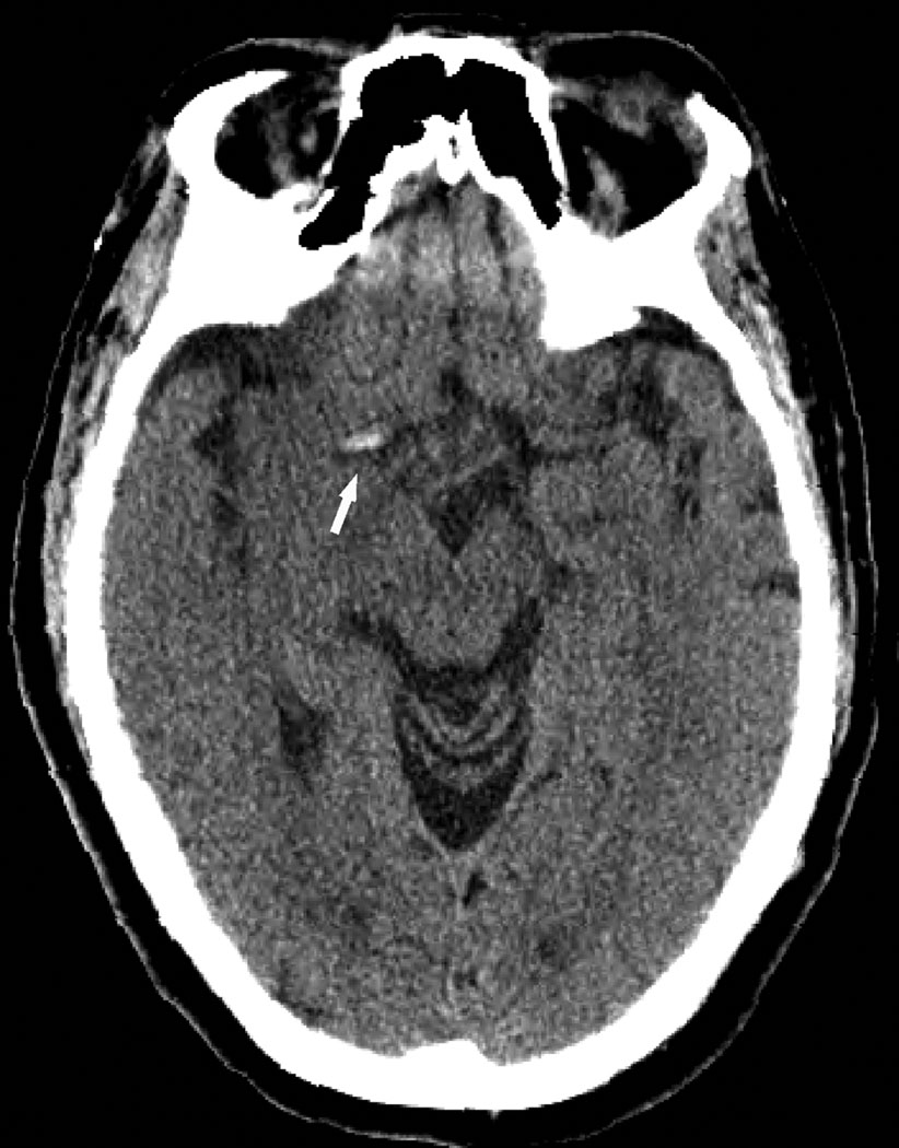
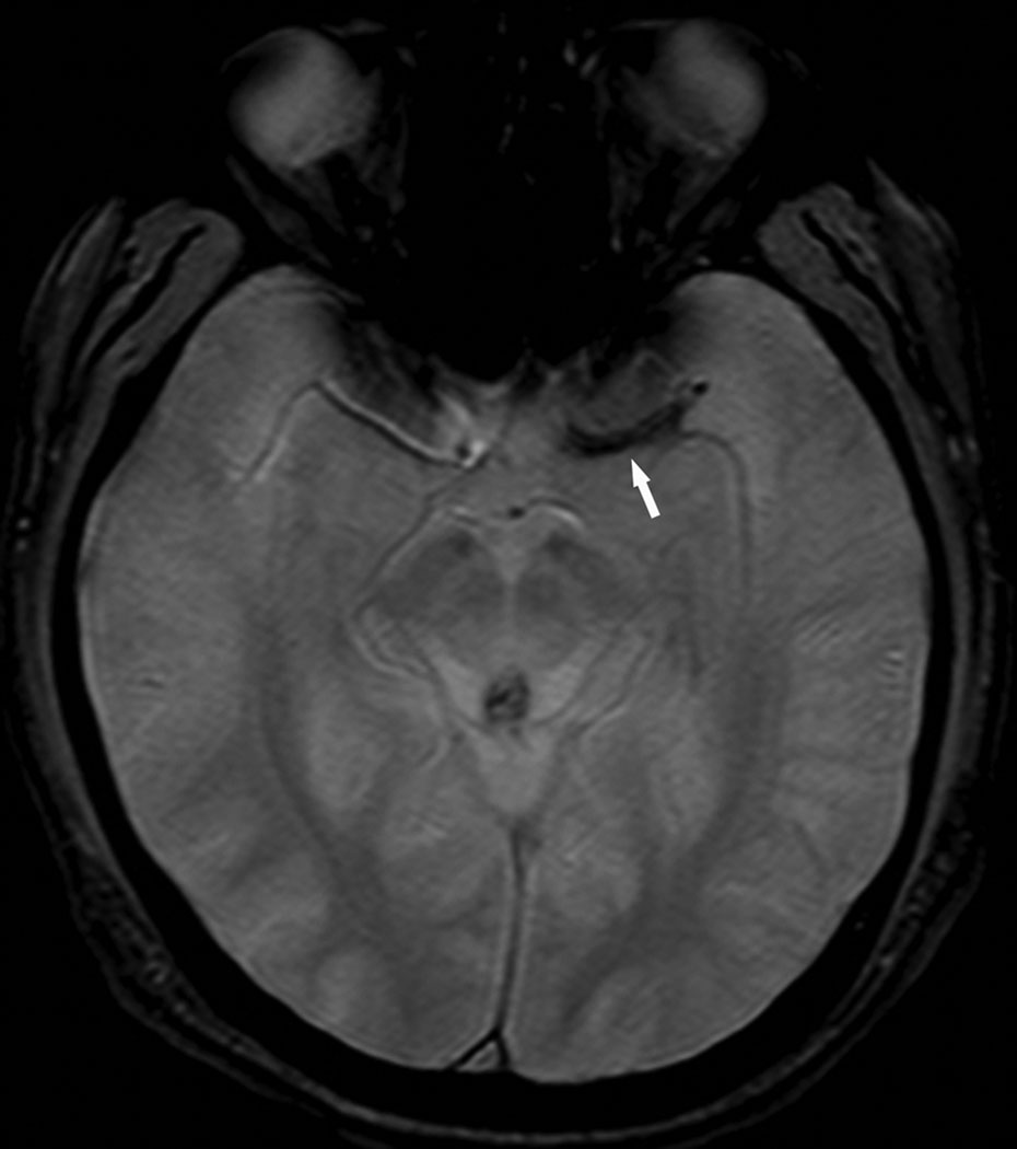
Background and purpose: The purpose of this study was to provide the first correlative study of the hyperdense middle cerebral artery sign (HMCAS) and gradient-echo MRI blooming artifact (BA) with pathology of retrieved thrombi in acute ischemic stroke.
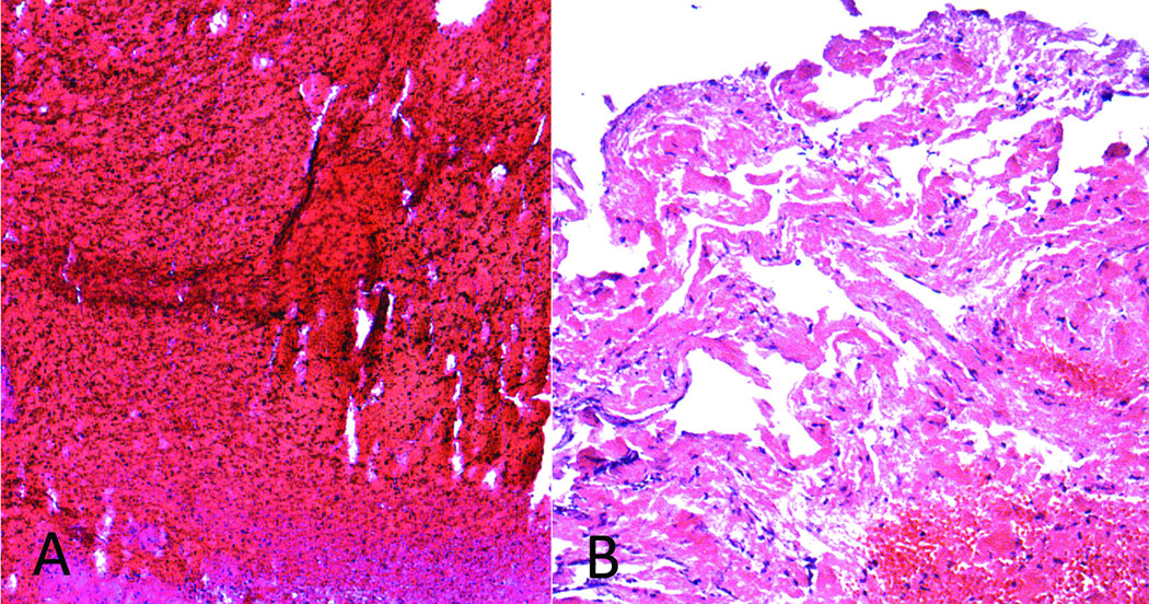
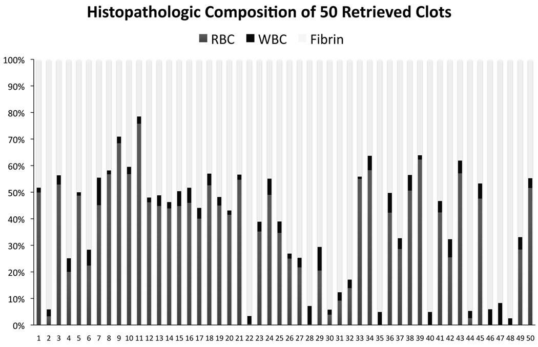
Methods: Noncontrast CT and gradient-echo MRI studies before mechanical thrombectomy in 50 consecutive cases of acute middle cerebral artery ischemic stroke were reviewed blinded to clinical and pathology data. Occlusions retrieved by thrombectomy underwent histopathologic analysis, including automated quantitative and qualitative rating of proportion composed of red blood cells (RBCs), white blood cells, and fibrin on microscopy of sectioned thrombi.
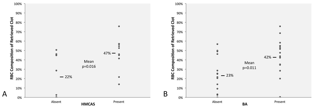
Results: Among 50 patients, mean age was 66 years and 48% were female. Mean (SD) proportion was 61% (±21) fibrin, 34% (±21) RBCs, and 4% (±2) white blood cells. Of retrieved clots, 22 (44%) were fibrin-dominant, 13 (26%) RBC-dominant, and 15 (30%) mixed. HMCAS was identified in 10 of 20 middle cerebral artery stroke cases with CT with mean Hounsfield Unit density of 61 (±8 SD). BA occurred in 17 of 32 with gradient-echo MRI. HMCAS was more commonly seen with RBC-dominant and mixed than fibrin-dominant clots (100% versus 67% versus 20%, P=0.016). Mean percent RBC composition was higher in clots associated with HMCAS (47% versus 22%, P=0.016). BA was more common in RBC-dominant and mixed clots compared with fibrin-dominant clots (100% versus 63% versus 25%, P=0.002). Mean percent RBC was greater with BA (42% versus 23%, P=0.011).
Conclusions: CT HMCAS and gradient-echo MRI BA reflect pathology of occlusive thrombus. RBC content determines appearance of HMCAS and BA, whereas absence of HMCAS or BA may indicate fibrin-predominant occlusive thrombi.
Conflict of interest statement
All authors were employed by the University of California (UC), which holds a patent on retriever devices for stroke, at the time of this work. The UC Regents received payments based on the clinical trial contracts for the number of subjects enrolled in the MR RESCUE multicenter clinical trial and the Concentric Merci Registry.
Dr. Liebeskind is a scientific consultant regarding trial design and conduct to Concentric Medical (modest) and CoAxia (modest).
Dr. Kidwell is Principal Investigator of the NIH-funded MR RESCUE trial (P50 NS044378).
Dr. Tateshima is a scientific advisor of Reverse Medical (modest), which makes a device to treat acute stroke.
Dr. Duckwiler is a medical advisor and stockholder of Concentric Medical.
Dr. Vinters is supported in part by the Daljit S. and Elaine Sarkaria Chair in Diagnostic Medicine.
Dr. Saver is a scientific consultant to AGA Medical (modest), Boehringer Ingelheim (modest), Bristol Myers Squibb (modest), CoAxia (modest), Concentric Medical (modest), Ev3 (modest), FibroGen (modest), ImaRx (modest), Sanofi Aventis (modest), and Talecris (modest). He receives support for editorial work in MedReviews (modest).
Figures
Comment in
-
Visualization of clot composition in ischemic stroke: do we get what we see?Stroke. 2011 May;42(5):1193-4. doi: 10.1161/STROKEAHA.110.612150. Epub 2011 Mar 10. Stroke. 2011. PMID: 21393600 No abstract available.
References
-
- Liebeskind DS. Reperfusion for acute ischemic stroke: Arterial revascularization and collateral therapeutics. Curr Opin Neurol. 2010;23:36–45. - PubMed
-
- Tissue plasminogen activator for acute ischemic stroke. The national institute of neurological disorders and stroke rt-pa stroke study group. N Engl J Med. 1995;333:1581–1587. - PubMed
-
- Gobin YP, Starkman S, Duckwiler GR, Grobelny T, Kidwell CS, Jahan R, Pile-Spellman J, Segal A, Vinuela F, Saver JL. Merci 1: A phase 1 study of mechanical embolus removal in cerebral ischemia. Stroke. 2004;35:2848–2854. - PubMed
-
- The penumbra pivotal stroke trial: Safety and effectiveness of a new generation of mechanical devices for clot removal in intracranial large vessel occlusive disease. Stroke. 2009;40:2761–2768. - PubMed
-
- Tomsick TA, Brott TG, Olinger CP, Barsan W, Spilker J, Eberle R, Adams H. Hyperdense middle cerebral artery: Incidence and quantitative significance. Neuroradiology. 1989;31:312–315. - PubMed
Publication types
MeSH terms
Substances
Grants and funding
LinkOut - more resources
Full Text Sources
Other Literature Sources
Medical
