. 2011 Mar;121(3):1154-62.
doi: 10.1172/JCI44198.

IL-21 is pivotal in determining age-dependent effectiveness of immune responses in a mouse model of human hepatitis B

Affiliations

IL-21 is pivotal in determining age-dependent effectiveness of immune responses in a mouse model of human hepatitis B

Jean Publicover et al. J Clin Invest. 2011 Mar.

Abstract

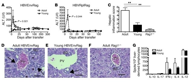
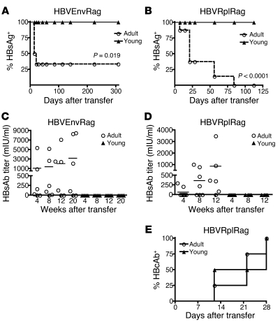
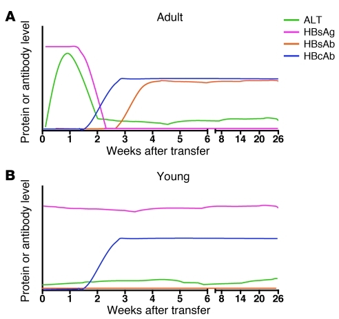
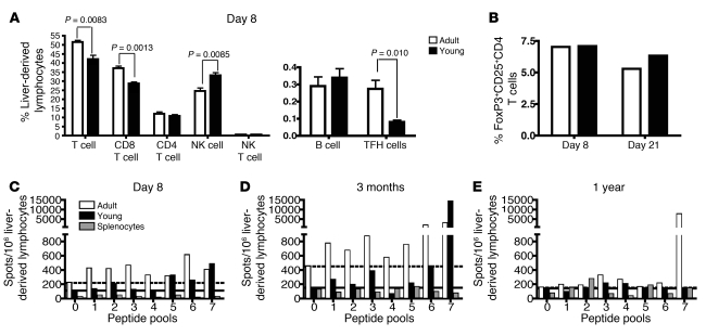
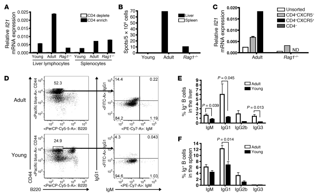
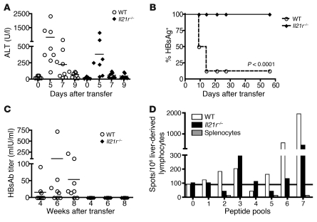
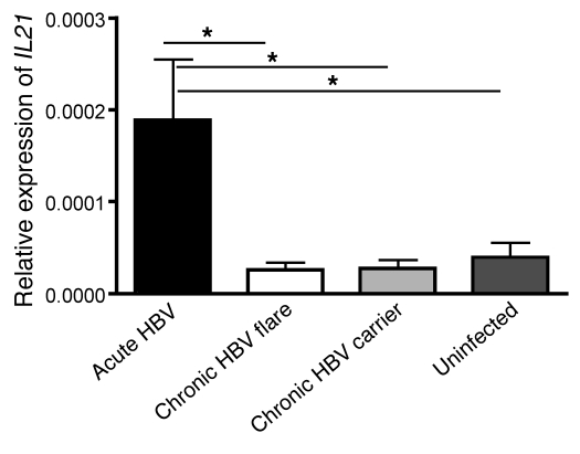
HBV is a noncytopathic hepadnavirus and major human pathogen that causes immune-mediated acute and chronic hepatitis. The immune response to HBV antigens is age dependent: viral clearance occurs in most adults, while neonates and children usually develop chronic infection and liver disease. Here, we characterize an animal model for HBV infection that recapitulates the key differences in viral clearance between early life and adulthood and find that IL-21 may be part of an effective primary hepatic immune response to HBV. In our model, adult mice showed higher HBV-dependent IL-21 production in liver, compared with that of young mice. Conversely, absence of the IL-21 receptor in adult mice resulted in antigen persistence akin to that of young mice. In humans, levels of IL-21 transcripts were greatly increased in blood samples from acutely infected adults who clear the virus. These observations suggest a different model for the dichotomous, age dependent outcome of HBV infection in humans, in which decreased IL-21 production in younger patients may hinder generation of crucial CD8+ T and B cell responses. These findings carry implications for therapeutic augmentation of immune responses to HBV and potentially other persistent liver viruses.


Figures

Figure 1
Figure 1. Transfer of adult splenocytes into young and adult HBVtgRag mice results in a difference in disease outcome.
Depiction of plasma ALT in young and adult (A) HBVEnvRag or (B) HBVRplRag mice after adoptive transfer of adult wild-type C57BL/6 mouse splenocytes. Error bars show mean ± SEM. Data are representative of at least 3 separate experiments where N ≥ 5 mice. Sections from formalin-fixed, paraffin-embedded liver tissue were taken 8 days after adoptive transfer of adult, syngeneic splenocytes and stained for H&E. (C) Composite score of necrosis, portal inflammation, and intraparenchymal inflammation as read by an unbiased pathologist. Error bars depict mean ± SEM; N ≥ 5 mice. Statistical significance was determined using the ANOVA with Tukey’s multiple comparison test. **P < 0.01. Representative liver H&E stains from (D) adult HBVEnvRag, (E) young HBVEnvRag, and (F) adult HBVtg-negative Rag1–/– mice. Arrows point to portal tract inflammation adjacent to the portal vein (PV) and bile duct (BD). Asterisks indicate necrotic hepatocytes. Scale bar: 30 mm. Statistical significance was determined using the unpaired 2-tailed t test. (G) IL-10, IL-17, IFN-γ, IL-4, and IL-12 cytokine levels were determined by ELISpot assay on liver-derived lymphocytes from adult or young HBVEnvRag mice 8 days after transfer of adult syngeneic splenocytes. Samples were pooled from n = 4 mice; error bars represent mean ± SEM from at least 3 separate wells.
Figure 2
Figure 2. Transfer of adult splenocytes into young and adult HBVtgRag mice results in a difference in HBsAg clearance and HBsAb responses.
Depiction of (A and B) the percentage of mice with detectable circulating HBsAg and (C and D) plasma HBsAb titer in young and adult (A and C) HBVEnvRag or (B and D) HBVRplRag mice after adoptive transfer of adult wild-type C57BL/6 mouse splenocytes. Error bars show mean ± SEM where N ≥ 5 mice. (E) Presence of HBcAb in plasma of adult or young HBVRplRag mice after adoptive transfer of adult, wild-type C57BL/6 mouse splenocytes (n = 4 mice). Statistical significance was determined using the unpaired 2-tailed t test. Data are representative of at least 3 separate experiments.
Figure 3
Figure 3. Patterns of serologic and molecular markers in this age-dependent model of primary HBV infection.
Typical levels of ALT, HBsAg, HBsAb, and HBcAb are shown in (A) adult HBVtgRag mice that received adult splenocytes by adoptive transfer and (B) young HBVtgRag mice that received adult splenocytes by adoptive transfer. The intensity of the responses, as a function of time after infection, is indicated schematically.
Figure 4
Figure 4. Adult HBVEnvRag recipient mice after adoptive transfer have more CD8+ T cells and TFH cells and elicit a more diverse and long-lived HBV-specific T cell response in the liver.
(A) The frequency of lymphocyte populations from the livers of adult and young HBVEnvRag mice 8 days after splenocyte transfer. TFH cells are defined as CD4+, CXCR5+, ICOS+ cells. B cells are defined as CD19+, B220+ cells. Error bars depict mean ± SEM; n = 4 mice. Statistical significance was determined using the unpaired 2-tailed t test. (B) Frequency of T regulatory cells in adult and young HBVEnvRag mouse liver-derived lymphocytes on days 8 and 21 after transfer of wild-type splenocytes. Samples were pooled from N ≥ 4 mice. Bars show percentages of CD4+-gated cells that are CD25+and FoxP3+. IFN-γ ELISpot results from young and adult HBVEnvRag mouse liver-derived lymphocytes (C) 8 days, (D) 3 months, or (E) 1 year after adoptive transfer. Cells were stimulated with HBV envelope peptide pools as described in Supplemental Figure 3 (pool 0 = no peptide). Dotted and solid lines indicate baseline IFN-γ levels for adult and young mice, respectively. A positive response was considered to be more than twice that of baseline. Lymphocytes were combined 1:1 with Rag1–/– splenocytes (to function as APCs); background IFN-γ levels from splenocytes are indicated by striped bars. Data are representative of at least 2 experiments; samples were pooled from N ≥ 4 mice.
Figure 5
Figure 5. Liver lymphocytes from adult HBVEnvRag mice after adoptive transfer produce more IL-21 from TFH cells and have increased numbers of IgG-expressing B cells.
(A) Il21 mRNA levels relative to those of GAPDH in CD4+ (CD4 enrich) and CD4 (CD4 deplete) fractions from liver-derived lymphocytes or splenocytes of adult or young HBVEnvRag or adult Rag1–/– mice 8 days after adoptive transfer of splenocytes. Error bars depict duplicate wells, samples were pooled from N ≥ 6 mice. (B) IL-21 protein expression determined by ELISpot on liver-derived lymphocytes and splenocytes 8 days after transfer. Samples were pooled from N ≥ 6 mice. (C) Il21 transcripts from unsorted liver lymphocytes, CXCR5CD4+ sorted cells, CXCR5+CD4+ sorted cells (not determined for Rag1–/– due to low cell number), and CXCR5CD4 cells from HBVEnvRag and Rag1–/– mice 8 days after transfer. Sorted cells were also CD19DAPI. Error bars depict triplicate wells; samples were pooled from N ≥ 4 mice. ND, not determined. (D) Frequency of B cells (plasmablasts and plasma cells) in adults and young HBVEnvRag mice 3 weeks after splenocyte transfer. The left plots show B220 versus CD44 expression on TCRβ populations, and the right plots show IgM versus IgG1 on TCRβ, CD44hi, B220lo populations. The percentage of IgG1-, IgG2b-, IgG3-, and IgM-expressing B cells from (E) liver-derived lymphocytes and (F) splenocytes. Error bars depict mean ± SEM; N ≥ 4 mice. Statistical significance was determined using the unpaired 2-tailed t test.
Figure 6
Figure 6. Adult HBVEnvRag mice fail to clear HBsAg, fail to produce HBsAb, and have a weaker and less diverse HBV-specific T cell response in the absence of IL-21R on transferred splenocytes.
Adult HBVEnvRag mice were adoptively transferred with either wild-type or IL-21R–deficient C57BL/6 splenocytes. (A) Mice were monitored for plasma ALT (no ALT differences were observed at later time points; data not shown). Horizontal bars indicate SEM. (B) The percentage of mice with detectable circulating HBsAg is shown. (C) Mice were monitored for HBsAb titer in the plasma (N ≥ 7 mice). Horizontal bars indicate SEM. (D) HBV-specific T cell responses at 2 months after transfer as measured by the IFN-γ ELISpot response of liver-derived lymphocytes stimulated with peptide pools; samples were pooled from N ≥ 4 mice. Statistical significance was determined using the unpaired 2-tailed t test.
Figure 7
Figure 7. Patients acutely infected with HBV express more IL21 mRNA in their PBMCs compared with patients with chronic HBV infection during actively flaring disease, inactive chronic HBV carriers, and healthy individuals.
IL21 mRNA relative to that of GAPDH in PBMCs taken from patients with acute HBV infection (high viral load, high ALT, IgM core Ab+, HBsAg+, and clinical history of exposure); patients with chronic HBV infection with a flare of disease (high ALT, high viral load, HBsAg+, and known history of chronic infection); patients with chronic HBV infection (low ALT, low viral load, and HBsAg+); and healthy, uninfected patients (low ALT and HBsAg) is shown. Error bars depict mean ± SEM; N ≥ 5. Statistical significance was determined using the ANOVA with Tukey’s post-hoc test. *P < 0.05.

Comment in

References

    1. Lee WM. Hepatitis B virus infection. N Engl J Med. 1997;337(24):1733–1745. doi: 10.1056/NEJM199712113372406. - DOI - PubMed
    1. Robinson WS. Hepatitis B virus and hepatitis D virus. In: Mandell GL, Bennett JE, Dolin R, eds.Principles and Practice of Infectious Diseases . 4th ed. New York, New York, USA: Churchill Livingstone; 1995:1406–1439.
    1. Ganem D, Prince AM. Hepatitis B virus infection--natural history and clinical consequences. N Engl J Med. 2004;350(11):1118–1129. doi: 10.1056/NEJMra031087. - DOI - PubMed
    1. Liang TJ. Hepatitis B: the virus and disease. Hepatology. 2009;49(5 suppl):S13–S21. doi: 10.1002/hep.22881. - DOI - PMC - PubMed
    1. Chisari FV, Isogawa M, Wieland SF. Pathogenesis of hepatitis B virus infection. Pathol Biol (Paris). 2010;58(4):258–266. - PMC - PubMed

Publication types