Clinical Trial
. 2011 Jun;31(6):1009-27.
doi: 10.1097/IAE.0b013e318217d739.

Randomized trial evaluating short-term effects of intravitreal ranibizumab or triamcinolone acetonide on macular edema after focal/grid laser for diabetic macular edema in eyes also receiving panretinal photocoagulation

Collaborators, Affiliations
Clinical Trial

Randomized trial evaluating short-term effects of intravitreal ranibizumab or triamcinolone acetonide on macular edema after focal/grid laser for diabetic macular edema in eyes also receiving panretinal photocoagulation

Diabetic Retinopathy Clinical Research Network et al. Retina. 2011 Jun.

Abstract

Purpose: To evaluate 14-week effects of intravitreal ranibizumab or triamcinolone in eyes receiving focal/grid laser for diabetic macular edema and panretinal photocoagulation.

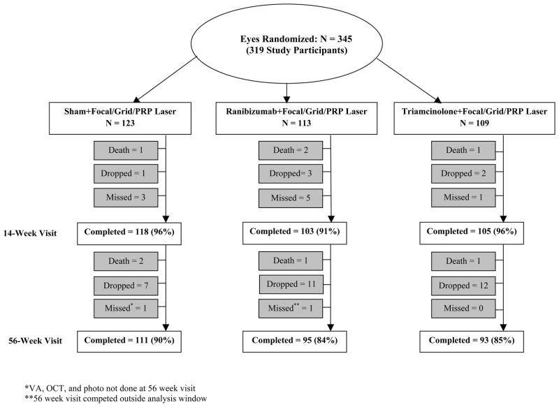
Methods: Three hundred and forty-five eyes with a visual acuity of 20/320 or better, center-involved diabetic macular edema receiving focal/grid laser, and diabetic retinopathy receiving prompt panretinal photocoagulation were randomly assigned to sham (n = 123), 0.5-mg ranibizumab (n = 113) at baseline and 4 weeks, and 4-mg triamcinolone at baseline and sham at 4 weeks (n = 109). Treatment was at investigator discretion from 14 weeks to 56 weeks.

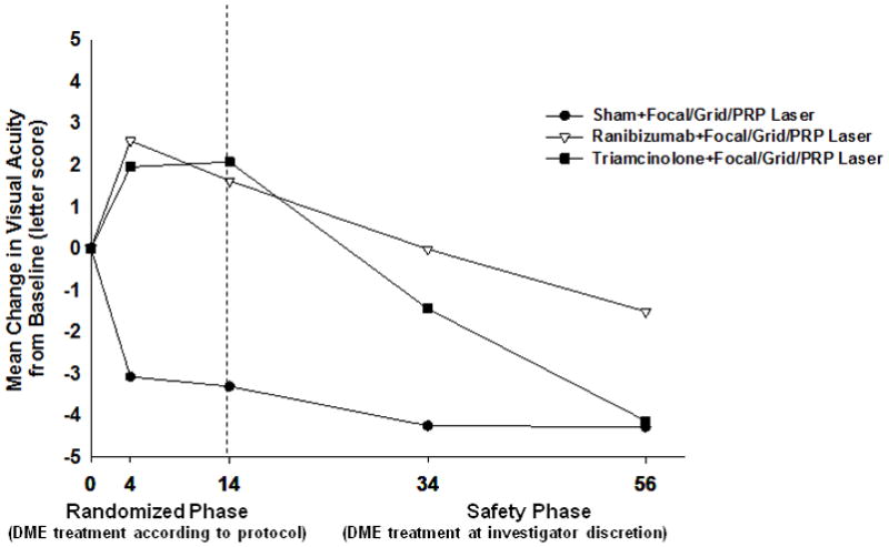
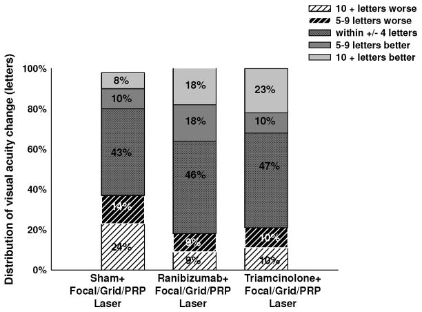
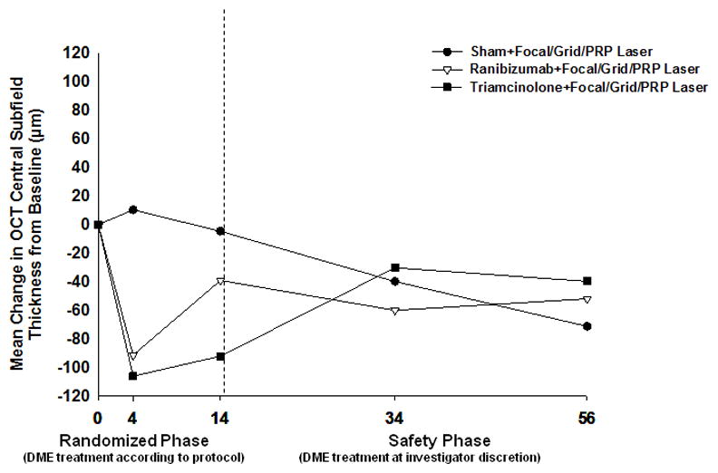
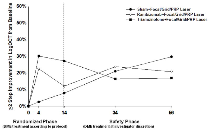
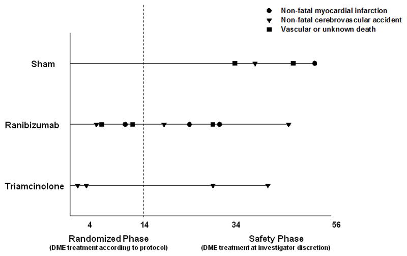
Results: Mean changes (±SD) in visual acuity letter score from baseline were significantly better in the ranibizumab (+1 ± 11; P < 0.001) and triamcinolone (+2 ± 11; P < 0.001) groups compared with those in the sham group (-4 ± 14) at the 14-week visit, mirroring retinal thickening results. These differences were not maintained when study participants were followed for 56 weeks for safety outcomes. One eye (0.9%; 95% confidence interval, 0.02%-4.7%) developed endophthalmitis after receiving ranibizumab. Cerebrovascular/cardiovascular events occurred in 4%, 7%, and 3% of the sham, ranibizumab, and triamcinolone groups, respectively.

Conclusion: The addition of 1 intravitreal triamcinolone injection or 2 intravitreal ranibizumab injections in eyes receiving focal/grid laser for diabetic macular edema and panretinal photocoagulation is associated with better visual acuity and decreased macular edema by 14 weeks. Whether continued long-term intravitreal treatment is beneficial cannot be determined from this study.


Conflict of interest statement

Financial Conflicts of Interest: A complete list of all DRCR.net investigator financial disclosures can be found at www.drcr.net

Figures

Figure 1
Figure 1
Completion of Follow-up for Study Eyes. Fourteen week completed visits include visits that occurred between 70 and 153 days (between 10 and 22 weeks) from randomization. Fifty-six week completed visits include visits that occurred between 315 and 468 days (between 45 and 67 weeks) from randomization. PRP=Panretinal photocoagulation.
Figure 2
Figure 2
Mean Change in Visual Acuity at Follow-up Visits. Values that were larger than ± 30 letters were assigned a value of 30. P values for difference in mean change in visual acuity from sham+focal/grid/PRP laser at the 14-week visit: ranibizumab+focal/grid/PRP laser <0.001 and triamcinolone+focal/grid/PRP laser groups <0.001. Fourteen week completed visits include visits that occurred between 70 and 153 days (between 10 and 22 weeks) from randomization. Fifty-six week completed visits include visits that occurred between 315 and 468 days (between 45 and 67 weeks) from randomization. PRP=Panretinal photocoagulation.
Figure 3
Figure 3
Distribution of Visual Acuity Change (letters) from Baseline to the 14-Week Visit. Fourteen week completed visits include visits that occurred between 70 and 153 days (between 10 and 22 weeks) from randomization. PRP=Panretinal photocoagulation.
Figure 4
Figure 4
Mean Change in Optical Coherence Tomography Central Subfield Retinal Thickening at Follow-up Visits. P values for difference in mean change in OCT central subfield retinal thickness from sham+focal/grid/PRP laser at the 14-week visit: ranibizumab+focal/grid/PRP laser = 0.01 and triamcinolone+focal/grid/PRP laser <0.001. Fourteen week completed visits include visits that occurred between 70 and 153 days (between 10 and 22 weeks) from randomization. Fifty-six week completed visits include visits that occurred between 315 and 468 days (between 45 and 67 weeks) from randomization. OCT = optical coherence tomography; PRP=Panretinal photocoagulation.
Figure 5
Figure 5
Two or More Step Improvement in the Logarithmic Transformation of Optical Coherence Tomography Central Subfield Thickness from Baseline. Fourteen week completed visits include visits that occurred between 70 and 153 days (between 10 and 22 weeks) from randomization. Fifty-six week completed visits include visits that occurred between 315 and 468 days (between 45 and 67 weeks) from randomization. logOCT = logarithmic transformation of optical coherence tomography calculated by taking the log base 10 of the ratio of the central subfield thickness divided by 200 and rounded to the nearest hundredth. DME=Diabetic macular edema; PRP=Panretinal photocoagulation.
Figure 6
Figure 6
Cardiovascular Events According to the Antiplatelet Trialists’ Collaboration* through 56-Week Visit. *Antiplatelet Trialists’ Collaboration. BMJ. 1994 Jan 8;308(6921):81–106. Non-fatal cerebrovascular accidents include ischemic, hemorrhagic or unknown. Vascular death includes any potential vascular or unknown cause. DME=Diabetic macular edema.

References

    1. The Diabetic Retinopathy Study Research Group. Preliminary report on effects of photocoagulation therapy. Am J Ophthalmol. 1976;81(4):383–96. - PubMed
    1. Ferris FL., 3rd How effective are treatments for diabetic retinopathy? JAMA. 1993;269(10):1290–1. - PubMed
    1. Myers SM. Macular edema after scatter laser photocoagulation for proliferative diabetic retinopathy. Am J Ophthalmol. 1980;90(2):210–6. - PubMed
    1. McDonald HR, Schatz H. Visual loss following panretinal photocoagulation for proliferative diabetic retinopathy. Ophthalmology. 1985;92(3):388–93. - PubMed
    1. Early Treatment Diabetic Retinopathy Study Research Group. Early photocoagulation for diabetic retinopathy. ETDRS report number 9. Ophthalmology. 1991;98(5 suppl):766–85. - PubMed

Publication types

MeSH terms