. 2011 Apr;13(4):394-401.
doi: 10.1038/ncb2207. Epub 2011 Mar 10.

Dynamics of ESCRT protein recruitment during retroviral assembly

Affiliations

Dynamics of ESCRT protein recruitment during retroviral assembly

Nolwenn Jouvenet et al. Nat Cell Biol. 2011 Apr.

Abstract

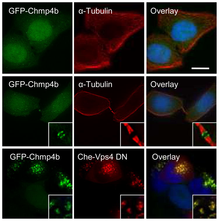
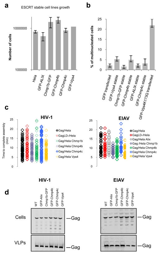
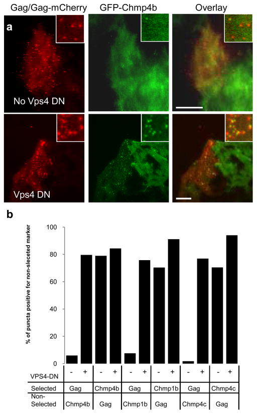
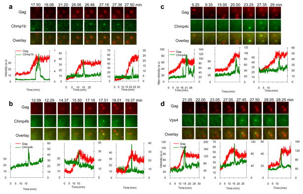
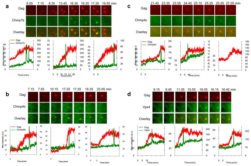
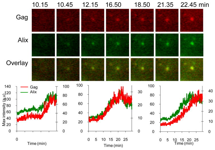
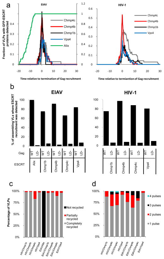
The ESCRT (endosomal sorting complex required for transport) complexes and associated proteins mediate membrane scission reactions, such as multivesicular body formation, the terminal stages of cytokinesis and retroviral particle release. These proteins are believed to be sequentially recruited to the site of membrane scission, and then complexes are disassembled by the ATPase Vps4A. However, these events have never been observed in living cells, and their dynamics are unknown. By quantifying the recruitment of several ESCRT and associated proteins during the assembly of two retroviruses, we show that Alix progressively accumulated at viral assembly sites, coincident with the accumulation of the main viral structural protein, Gag, and was not recycled after assembly. In contrast, ESCRT-III and Vps4A were transiently recruited only when the accumulation of Gag was complete. These data indicate that the rapid and transient recruitment of proteins that act late in the ESCRT pathway and carry out membrane fission is triggered by prior and progressive accumulation of proteins that bridge viral proteins and the late-acting ESCRT proteins.


Conflict of interest statement

The authors declare no competing financial interests.

Figures

Figure 1
Figure 1. Characterization of the GFP-Chmp4b-expressing cell clone
Hela cells stably expressing GFP-Chmp4b (green) were fixed and stained with anti-α-tubulin antibodies (red) and with DAPI (blue). Images show the distribution of GFP-Chmp4b in interphase cells (top panels) and in telophase cells (middle panels). Alternatively, cells were transfected with mCherry-Vps4-DN (bottom panels), fixed 18 hours post-transfection and stained with DAPI (blue). Samples were observed with an epifluorescence microscope. Deconvolved optical sections acquired at the center of the vertical dimension of the cell are shown. Expanded views are shown in insets. The scale bars represent 10μm.
Figure 2
Figure 2. Effect of stably expressed GFP fused ESRCT proteins on cell proliferation, cytokinesis and virion assembly and release
a. The stable expression of GFP-tagged ESCRT proteins does not affect cell proliferation. 105 cells were plated into each well of a 24-well plate, harvested and counted 48h later. Error bars indicate s.d from 3 independent experiments. b. Stable expression of GFP-tagged ESCRT proteins does not disrupt cytokinesis. Hela cells stably expressing GFP-tagged ESCRT proteins or transfected with GFP or GFP-Vps4-K173Q were fixed, stained with both anti-α-tubulin antibodies and DAPI, and scored for multinucleated cells. 300 cells from 3 independent experiments were analyzed for the presence of more than one nucleus per cell for each factor. Error bars indicate s.d. c. Kinetics of HIV-1 and EIAV assembly were not affected in the cell lines stably expressing GFP-tagged ESCRT proteins. The plots show the time to complete assembly for individual HIV-1 and EIAV VLPs, including wild-type and late domain mutants (LD-) Gag proteins, in unmodified Hela cells, or cell lines expressing the indicated GFP-ESCRT proteins lines. Each symbol represents an individual VLP. The time to complete assembly was defined for each VLP as the interval between the points of inflection on plots of fluorescence intensity versus time. d. The stable cell lines stably expressing GFP-tagged ESCRT proteins support the release of HIV-1 and EIAV Gag VLPs. Western blot analysis (LICOR) of Hela cells and Hela cells stably expressing GFP-tagged ESCRT transfected with HIV-1 (left panel) or EIAV (right panel) Gag. Samples were probed with an anti-p24 monoclonal antibody for HIV-1 or anti-EIAV horse serum. Fig. S6 is showing the corresponding unprocessed western blots.
Figure 3
Figure 3. Catalytically inactive Vps4A increases localization of stably expressed GFP tagged ESCRT-III proteins at sites of HIV-1 assembly
a. Hela cells stably expressing GFP-Chmp4b (green) were transfected with HIV-1 Gag/Gag-mCherry (red), in the absence (top panel) or presence (bottom panel) of Vps4A- DN. Cells were fixed 24 hours later and observed with a TIR-FM microscope. Expanded views are shown in insets. The scale bar represents 10μm. b. Quantification of the co-localization between VLPs and puncta of ESCRT proteins. Hela cells stably expressing GFP-fused ESCRT-III proteins were transfected with Gag/Gag-mCherry, in the absence(−) or presence (+) of Vps4A-DN, as indicated. Cells were observed under TIR-FM at 18 hours post-transfection and the co-localization between puncta of Gag-mCherry and puncta of GFP was quantified by randomly selecting puncta of one marker (selected) and then enumerating what percentage of these puncta were coincident with puncta of the other, non selected marker.
Figure 4
Figure 4. Imaging Chmp1b, Chmp4b, Chmp4c and Vps4A recruitment during HIV-1 Gag assembly
Hela cells stably expressing Chmp1b-GFP (a), GFP-Chmp4b (b), GFP-Chmp4c (c) or GFP-Vps4A (d) were transfected with HIV-1 Gag/Gag-mCherry and observed under TIR-FM beginning at 6 hours post-transfection. Each set of images illustrates the recruitment of GFP-labeled ESCRT proteins (green) during the genesis of an individual VLP (red). The time after the commencement of observation is given in minutes:seconds. Fields are 2.5x2.5μm. Plots of fluorescence intensity in arbitrary units (a.u) over time for the GFP-ESCRT protein (green, right axis) and Gag-mCherry signals (red, left axis) associated with the assembly of 3 individual VLPs are shown.
Figure 5
Figure 5. Imaging Chmp1b, Chmp4b, Chmp4c and Vps4A recruitment during EIAV Gag assembly
Hela cells stably expressing Chmp1b-GFP (a), GFP-Chmp4b (b), GFP-Chmp4c (c) or GFP-Vps4A (d) were transfected with EIAV Gag/Gag-mCherry and observed under TIR-FM beginning at 6 hours post-transfection. Each set of images illustrates the recruitment of GFP-labeled ESCRT proteins during the genesis of an individual VLP. The time after the commencement of observation is given in minutes:seconds. Fields are 2.5x2.5μm. Plots of fluorescence intensity in arbitrary units (a.u) over time for the GFP-ESCRT protein (green, right axis) and Gag-mCherry signals (red, left axis) associated with the assembly of 3 individual VLPs are shown, the left chart in panels a and d correspond to the microscopic images shown above.
Figure 6
Figure 6. Imaging Alix recruitment during EIAV Gag assembly
Hela cells stably expressing GFP-Alix were transfected with EIAV Gag/Gag-mCherry and observed under TIR-FM beginning at 6 hours post-transfection. Each set of images illustrates the recruitment of GFP-labeled ESCRT proteins during the genesis of an individual VLP. The time after the commencement of observation is given in minutes:seconds. Fields are 2.5x2.5μm. Plots of fluorescence intensity in arbitrary units (a.u) over time for the GFP-ESCRT protein (green, right axis) and Gag-mCherry signals (red, left axis) associated with the assembly of 3 individual VLPs are shown, the left chart correspond to the microscopic images shown above.
Figure 7
Figure 7. Dynamics and pattern of ESCRT proteins recruitment during retroviral assembly
a. The fraction of EIAV (left panel) and HIV-1 (right panel) VLPs at which GFP-ESCRT-protein was detectable is plotted as a function of time. For this analysis, T=0 was set as the point at which Gag recruitment to each VLP reached a plateau. b. Proportion (%) of VLP assembly events for which the recruitment of GFP-tagged ESCRT proteins was detected. Hela cells stably expressing GFP-tagged ESCRT proteins were transfected with wild-type or L-domain mutant (LD−) HIV-1 (left panel) or EIAV (right panel) Gag/Gag-mCherry. Cells were observed live under TIR-FM beginning at 6 hours post-transfection, for a period of 25 to 50 minutes. c. Quantification of the number of pulses of ESCRT protein recruitment (percentage of VLPs for which each behavior is observed) during HIV-1 and EIAV VLP assembly d. Removal of the GFP-ESCRT proteins from sites of HIV-1 and EIAV assembly. ESCRT-III and Vps4 proteins are generally completely recycled (and signified by the GFP signal at VLP assembly sites returning to baseline levels following the pulse), but in some cases, the proteins appear to be only partially recycled (e.g. Fig. S8, left panel). Alix is not recycled (i.e the GFP signal remains at a plateau after reaching its maximum, see Fig 3c). The percentage of VLPs showing each behavior is plotted.

Comment in

References

    1. McDonald B, Martin-Serrano J. No strings attached: the ESCRT machinery in viral budding and cytokinesis. J Cell Sci. 2009;122:2167–2177. - PMC - PubMed
    1. Wollert T, et al. The ESCRT machinery at a glance. J Cell Sci. 2009;122:2163–2166. - PMC - PubMed
    1. Bieniasz PD. Late budding domains and host proteins in enveloped virus release. Virology. 2006;344:55–63. - PubMed
    1. Morita E, Sundquist WI. Retrovirus budding. Annu Rev Cell Dev Biol. 2004;20:395–425. - PubMed
    1. Williams RL, Urbe S. The emerging shape of the ESCRT machinery. Nat Rev Mol Cell Biol. 2007;8:355–368. - PubMed

Publication types

MeSH terms

Substances