

Baseline mean deviation and rates of visual field change in treated glaucoma patients
- PMID: 21394112
- PMCID: PMC3171256
- DOI: 10.1038/eye.2011.33
Baseline mean deviation and rates of visual field change in treated glaucoma patients
Abstract
Purpose: To evaluate the relationships between baseline visual field (VF) mean deviation (MD) and subsequent progression in treated glaucoma.
Methods: Records of patients seen in a glaucoma practice between 1999 and 2009 were reviewed. Patients with glaucomatous optic neuropathy, baseline VF damage, and ≥8 SITA-standard 24-2 VF were included. Patients were divided into tertiles based upon baseline MD. Automated pointwise linear regression determined global and localized rates (decibels (dB) per year) of change. Progression was defined when two or more adjacent test locations in the same hemifield showed a sensitivity decline at a rate of >1.0 dB per year, P<0.01.
Results: For mild, moderate, and severe groups, progression was noted in 29.5, 31.2, and 26.0% of eyes (P=0.50) and global rates of VF change of progressing eyes were -1.3±1.2, -1.01±0.7, and -0.9±0.5 dB/year (P=0.09, analysis of variance). Within these groups, intraocular pressure (IOP) in stable vs progressing eyes were 15.5±3.3 vs 17.0±3.1 (P<0.01), 15.4±3.3 vs 15.9±2.5 (P=0.28), and 14.0±2.8 vs 14.8±2.3 mm Hg (P=0.07). More glaucoma filtering surgeries were performed in eyes with worse MD. There was no significant difference between groups regarding their risk of progression in both univariate (P=0.50) and multivariate (P=0.26) analyses adjusting for differences in follow-up IOP.
Conclusions: After correcting for differences in IOP in treated glaucoma patients, we did not find a relationship between the rate of VF change (dB per year) and the severity of the baseline VF MD. This finding may have been due to more aggressive IOP lowering in eyes with more severe disease. Eyes with lower IOP progressed less frequently across the spectrum of VF loss.
Figures
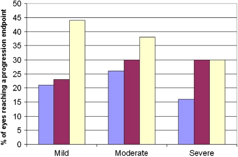
 , 1st IOP tertile;
  , 2nd IOP tertile;
  , 3rd IOP tertile. Mean IOP values for each tertile: mild: <14.68, 14.68–17.45, and 17.45–23.81 mm Hg; moderate: <14.50, 14.50–16.80, and 16.80–22.81 mm Hg; severe: <13.31, 13.31–15.35, and 15.35–21.58 mm Hg.
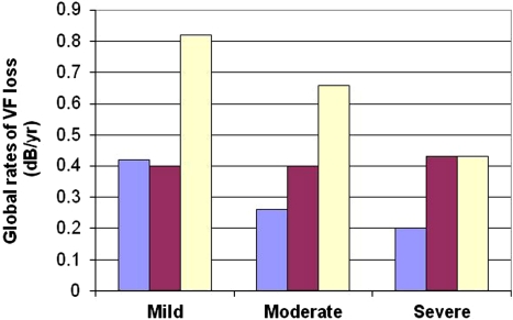
 , 1st IOP tertile;
  , 2nd IOP tertile;
  , 3rd IOP tertile. Mean IOP values for each tertile: mild: <14.68, 14.68–17.45, and 17.45–23.81 mm Hg; moderate: <14.50, 14.50–16.80, and 16.80–22.81 mm Hg; severe: <13.31, 13.31–15.35, and 15.35–21.58 mm Hg.
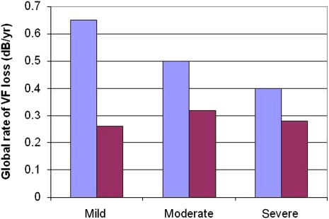
 , 1st IOP tertile;
  , 2nd IOP tertile.
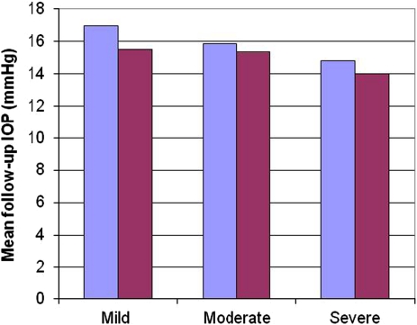
 , 1st IOP tertile;
  , 2nd IOP tertile.
References
-
- Kass MA, Heuer DK, Higginbotham EJ, Johnson CA, Keltner JL, Miller JP, et al. The Ocular Hypertension Treatment Study: a randomized trial determines that topical ocular hypotensive medication delays or prevents the onset of primary open-angle glaucoma. Arch Ophthalmol. 2002;120:701–713. - PubMed
-
- Heijl A, Leske MC, Bengtsson B, Hyman L, Bengtsson B, Hussein M. Early Manifest Glaucoma Trial Group. Reduction of intraocular pressure and glaucoma progression: results from the Early Manifest Glaucoma Trial. Arch Ophthalmol. 2002;120:1268–1279. - PubMed
-
- AGIS Investigators The Advanced Glaucoma Intervention Study (AGIS): 7. The relationship between control of intraocular pressure and visual field deterioration. Am J Ophthalmol. 2000;130:490–491. - PubMed
-
- Lichter PR, Musch DC, Gillespie BW, Guire KE, Janz NK, Wren PA, et al. CIGTS Study Group. Interim clinical outcomes in the Collaborative Initial Glaucoma Treatment Study comparing initial treatment randomized to medications or surgery. Ophthalmology. 2001;108:1943–1953. - PubMed
-
- Miglior S, Torri V, Zeyen T, Pfeiffer N, Vaz JC, Adamsons I. Intercurrent factors associated with the development of open-angle glaucoma in the European glaucoma prevention study. Am J Ophthalmol. 2007;144 (2:266–275. - PubMed
Publication types
MeSH terms
LinkOut - more resources
Full Text Sources
Medical
Miscellaneous
