

Enhancing the response of CALUX and CAFLUX cell bioassays for quantitative detection of dioxin-like compounds
- PMID: 21394221
- PMCID: PMC3049243
- DOI: 10.1007/s11426-010-0142-8
Enhancing the response of CALUX and CAFLUX cell bioassays for quantitative detection of dioxin-like compounds
Abstract
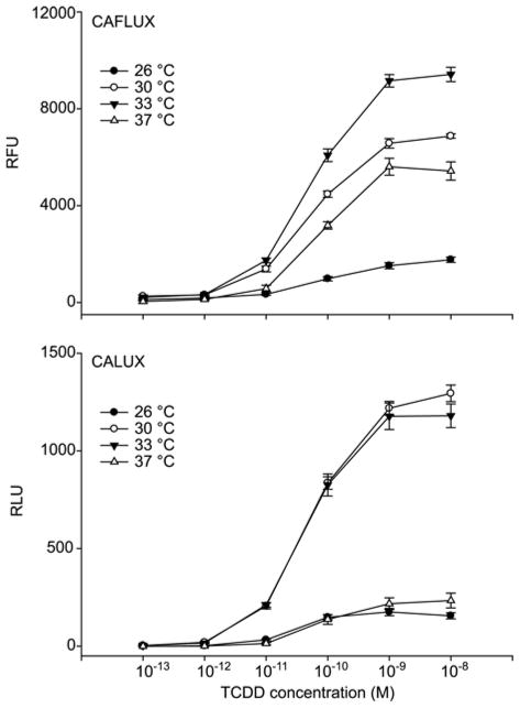
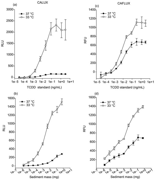
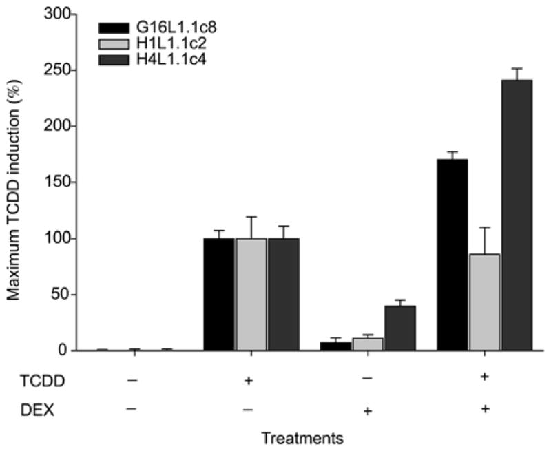
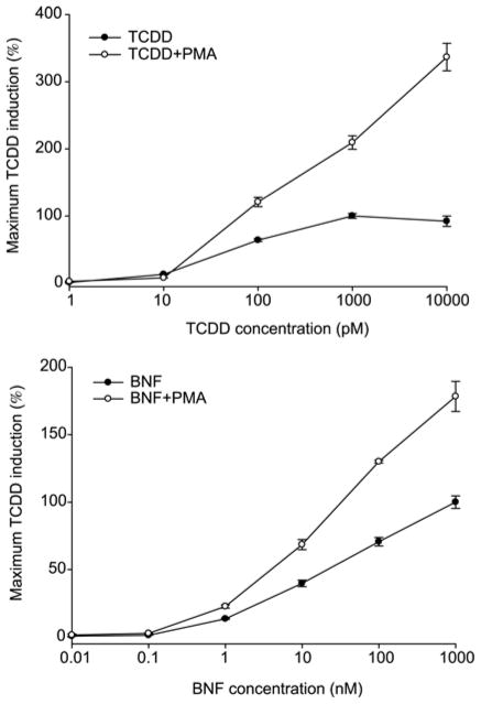
Reporter genes produce a protein product in transfected cells that can be easily measured in intact or lysed cells and they have been extensively used in numerous basic and applied research applications. Over the past 10 years, reporter gene assays have been widely accepted and used for analysis of 2,3,7,8-tetrachlorodibenzo-p-dioxin and related dioxin-like compounds in various types of matrices, such as biological, environmental, food and feed samples, given that high-resolution instrumental analysis techniques are impractical for large-scale screening analysis. The most sensitive cell-based reporter gene bioassay systems developed are the mechanism-based CALUX (Chemically Activated Luciferase Expression) and CAFLUX (Chemically Activated Fluorescent Expression) bioassays, which utilize recombinant cell lines containing stably transfected dioxin (AhR)-responsive firefly luciferase or enhanced green fluorescent protein (EGFP) reporter genes, respectively. While the current CALUX and CAFLUX bioassays are very sensitive, increasing their lower limit of sensitivity, magnitude of response and dynamic range for chemical detection would significantly increase their utility, particularly for those samples that contain low levels of dioxin-like HAHs (i.e., serum). In this study, we report that the addition of modulators of cell signaling pathways or modification of cell culture conditions results in significant improvement in the magnitude and overall responsiveness of the existing CALUX and CAFLUX cell bioassays.
Figures
References
-
- Behnisch PA, Hosoe K, Sakai S. Bioanalytical screening methods for dioxins and dioxin-like compounds a review of bioassay/biomarker technology. Environ Int. 2001;27:413–439. - PubMed
-
- Hahn ME. Biomarkers and bioassays for detecting dioxin-like compounds in the marine environment. Sci Total Environ. 2002;289:49–69. - PubMed
-
- Denison MS, Zhao B, Baston DS, Clark GC, Murata H, Han DH. Recombinant cell bioassay systems for the detection and relative quantitation of halogenated dioxins and related chemicals. Talanta. 2004;63:1123–1133. - PubMed
-
- Whyte JJ, Schmitt CJ, Tillitt DE. The H4IIE cell bioassay as an indicator of dioxin-like chemicals in wildlife and the environment. Crit Rev Toxicol. 2004;34:1–83. - PubMed
-
- Harrison RO, Eduliee GH. Immunochemical analysis for dioxins-progress and prospects. Sci Total Environ. 1999;239:1–18. - PubMed
Grants and funding
LinkOut - more resources
Full Text Sources
Research Materials
Miscellaneous