

An in vivo comparison of hip structure analysis (HSA) with measurements obtained by QCT
- PMID: 21394495
- PMCID: PMC3261404
- DOI: 10.1007/s00198-011-1578-1
An in vivo comparison of hip structure analysis (HSA) with measurements obtained by QCT
Abstract
Summary: In a population of elderly women, bone cross-sectional area (CSA), cross-sectional moment of inertia (CSMI), section modulus (Z), femoral neck axis length (FNAL), and width measured with hip structure analysis (HSA) on dual-energy x-ray absorptiometry (DXA) images in the femoral neck and trochanteric regions are highly correlated to quantitative computed tomography (QCT) measurements.
Introduction: HSA is a method of obtaining measurements of proximal femur structure using 2D DXA technology. This study was designed to examine the correlations between HSA measurements and 3D QCT.
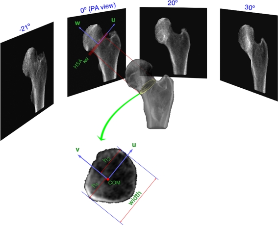
Methods: Forty-one women (mean age, 82.8 ± 2.5 years) were measured using DXA and a 64-slice CT scanner (1 mm slice thickness, 0.29 mm in plane resolution). HSA parameters were calculated at the narrow neck (NN) and trochanteric (IT) regions on the DXA image. These regions were then translated to anatomically equivalent regions on the QCT dataset by co-registering the DXA image and QCT dataset using four DXA images acquired at different angles.
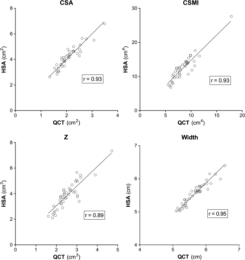
Results: At the NN and IT regions, high linear correlations were measured between HSA and QCT for CSA r = 0.95 and 0.93, CSMI r = 0.94 and 0.93, and Z r = 0.93 and 0.89, respectively. All correlations were highly significant (p < 0.001), but there were differences in slope and offset between the two techniques, at least in part due to differences in calibration between the two techniques. FNAL and width of the bone at the NN and IT regions, physical measurements independent of the calibration, were highly correlated (r = 0.90-0.95, p < 0.001) and had slopes close to 1.0 (range, 0.978 to 1.003).
Conclusion: CSA, CSMI, Z, FNAL, and width measured by HSA correlate highly to high-resolution QCT.
Figures


References
-
- Beck TJ, Looker AC, Ruff CB, Sievanen H, Wahner HW. Structural trends in the aging femoral neck and proximal shaft: analysis of the Third National Health and Nutrition Examination Survey dual-energy X-ray absorptiometry data. J Bone Miner Res. 2000;15:2297–2304. doi: 10.1359/jbmr.2000.15.12.2297. - DOI - PubMed
Publication types
MeSH terms
LinkOut - more resources
Full Text Sources
