. 2011 Apr;34(2):141-53.
doi: 10.1007/s13402-011-0019-7. Epub 2011 Mar 11.

Phospho-proteomic analysis of mantle cell lymphoma cells suggests a pro-survival role of B-cell receptor signaling

Affiliations

Phospho-proteomic analysis of mantle cell lymphoma cells suggests a pro-survival role of B-cell receptor signaling

Chiara Pighi et al. Cell Oncol (Dordr). 2011 Apr.

Abstract

Background: Mantle cell lymphoma (MCL) is currently an incurable entity, and new therapeutic approaches are needed. We have applied a high-throughput phospho-proteomic technique to MCL cell lines to identify activated pathways and we have then validated our data in both cell lines and tumor tissues.

Methods: PhosphoScan analysis was performed on MCL cell lines. Results were validated by flow cytometry and western blotting. Functional validation was performed by blocking the most active pathway in MCL cell lines.

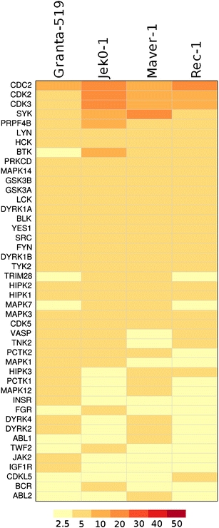
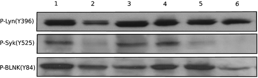
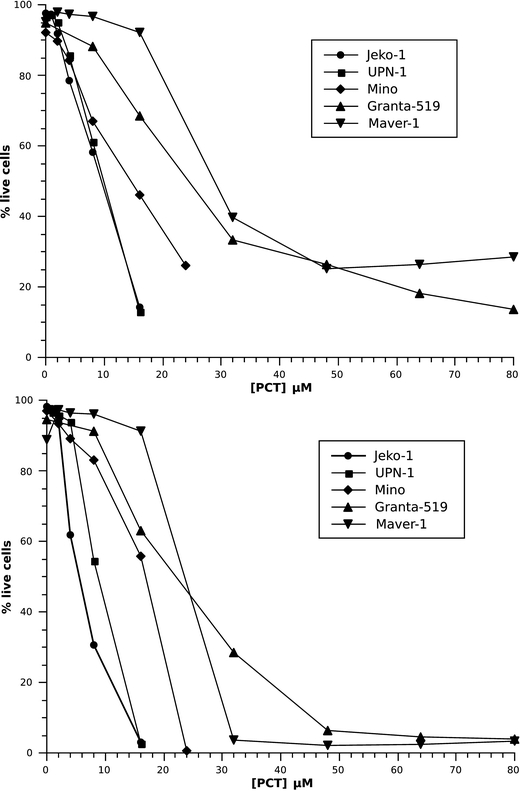
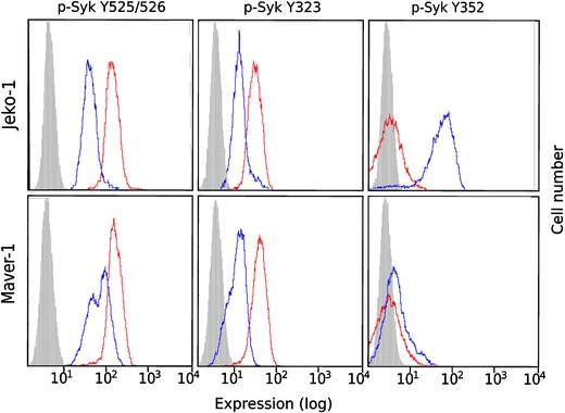
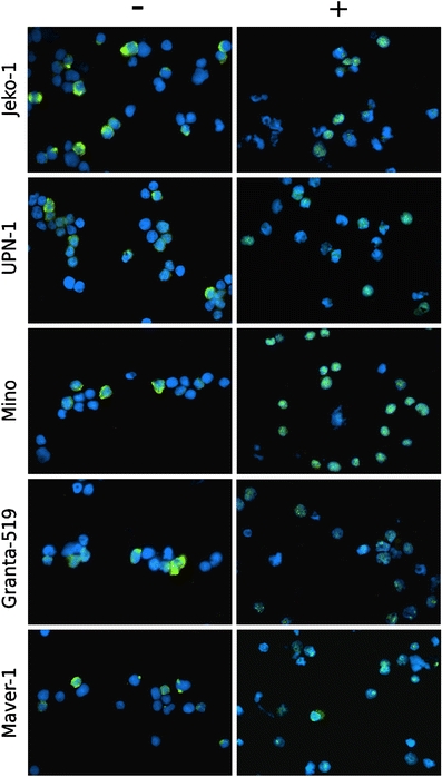
Results: PhosphoScan identified more than 300 tyrosine-phosporylated proteins, among which many protein kinases. The most abundant peptides belonged to proteins connected with B-cell receptor (BCR) signaling. Active BCR signaling was demonstrated by flow cytometry in MCL cells and by western blotting in MCL tumor tissues. Blocking BCR signaling by Syk inhibitor piceatannol induced dose/time-dependent apoptosis in MCL cell lines, as well as several modifications in the phosphorylation status of BCR pathway members and a collapse of cyclin D1 protein levels.

Conclusion: Our data support a pro-survival role of BCR signaling in MCL and suggest that this pathway might be a candidate for therapy. Our findings also suggest that Syk activation patterns might be different in MCL compared to other lymphoma subtypes.


Figures

Fig. 1
Fig. 1
List of protein kinases identified by the PhosphoScan approach, ranked by overall abundance in the four MCL cell lines analyzed. Color intensity proportional to the number of peptides (see figure for color corresponding to the average number of peptides per cell line)
Fig. 2
Fig. 2
Simplified diagram showing some of the identified BCR pathway members. In red, activating members; in green, inhibiting members; for some members the precise final effect is not clear; in grey, other proteins known to be present but not identified. Full arrows: direct interaction/activation; dotted arrows: indirect interaction/activation. Data derived from Kyoto Encyclopedia of Genes and Genomes (KEGG) pathway [37] and from published literature. KEGG is a widely used annotated database of pathways, ligands and genes (http://www.genome.jp/kegg/)
Fig. 3
Fig. 3
Analysis of basal levels of phosphorylated Syk and Blnk residues by flow cytometry. In grey, isotypic control; in red, basal levels. On the X axis, arbitrary fluorescence units (log scale); on the Y axis, cell count
Fig. 4
Fig. 4
Western blotting analysis of MCL tissues. The presence of phospho-Syk (Y525), phospho-Lyn (Y396) and phospho-Blnk (Y84) is shown in six MCL tumor tissues. Cases 1, 2, 5 and 6 were classical MCL, while cases 3 and 4 were blastoid variants
Fig. 5
Fig. 5
Induction of apoptosis in MCL cell lines. Upper panel shows the percentage of live cells (Y axis) in function of the piceatannol concentration (X axis) at 24 h. Lower panel shows the same variables at 48 h of treatment. Annexin V staining was used to discriminate apoptotic cells
Fig. 6
Fig. 6
Modification of Syk phosphorylation profile following piceatannol treatment. After treatment, phosphorylation of residues Y525 and Y323 is reduced, while that of residue Y352 is increased. Red, untreated cells; blue, treated cells; solid grey, isotypic control
Fig. 7
Fig. 7
Localization of phospho-Syk following piceatannol treatment. P-Syk Y525 (shown in green pseudo-color) disappears from the cytoplasm and appears in the nucleus after treatment in all cell lines. Nuclei counter stained in blue pseudo-color (DAPI staining)

References

    1. Swerdlow SH, Campo E, Seto M, Muller-Hermelink HK. In: Mantle cell lymphoma, in: WHO classification of tumours of haematopoietic and lymphoid tissues. Swerdlow SH, Campo E, Harris NL, Jaffe ES, Pileri S, Stein H, Thiele J, Vardiman J, editors. Lyon: IARC Press; 2008.
    1. Hofmann WK, de Vos S, Tsukasaki K, Wachsman W, Pinkus GS, Said JW, Koeffler HP. Altered apoptosis pathways in mantle cell lymphoma detected by oligonucleotide microarray. Blood. 2001;98:787–794. doi: 10.1182/blood.V98.3.787. - DOI - PubMed
    1. Martinez N, Camacho FI, Algara P, Rodriguez A, Dopazo A, Ruiz-Ballesteros E, Martin P, Martinez-Climent JA, Garcia-Conde J, Menarguez J, Solano F, Mollejo M, Piris MA. The molecular signature of mantle cell lymphoma reveals multiple signals favoring cell survival. Cancer Res. 2003;63:8226–8232. - PubMed
    1. Ortega-Paino E, Fransson J, Ek S, Borrebaeck CA. Functionally associated targets in mantle cell lymphoma as defined by DNA microarrays and RNA interference. Blood. 2008;111:1617–1624. doi: 10.1182/blood-2007-02-068791. - DOI - PubMed
    1. Pinyol M, Bea S, Pla L, Ribrag V, Bosq J, Rosenwald A, Campo E, Jares P. Inactivation of RB1 in mantle-cell lymphoma detected by nonsense-mediated mRNA decay pathway inhibition and microarray analysis. Blood. 2007;109:5422–5429. doi: 10.1182/blood-2006-11-057208. - DOI - PubMed

Publication types

MeSH terms