Review
. 2011 Mar 25;23(12):H41-56.
doi: 10.1002/adma.201003963. Epub 2011 Mar 10.

Hyaluronic acid hydrogels for biomedical applications

Affiliations
Review

Hyaluronic acid hydrogels for biomedical applications

Jason A Burdick et al. Adv Mater. .

Abstract

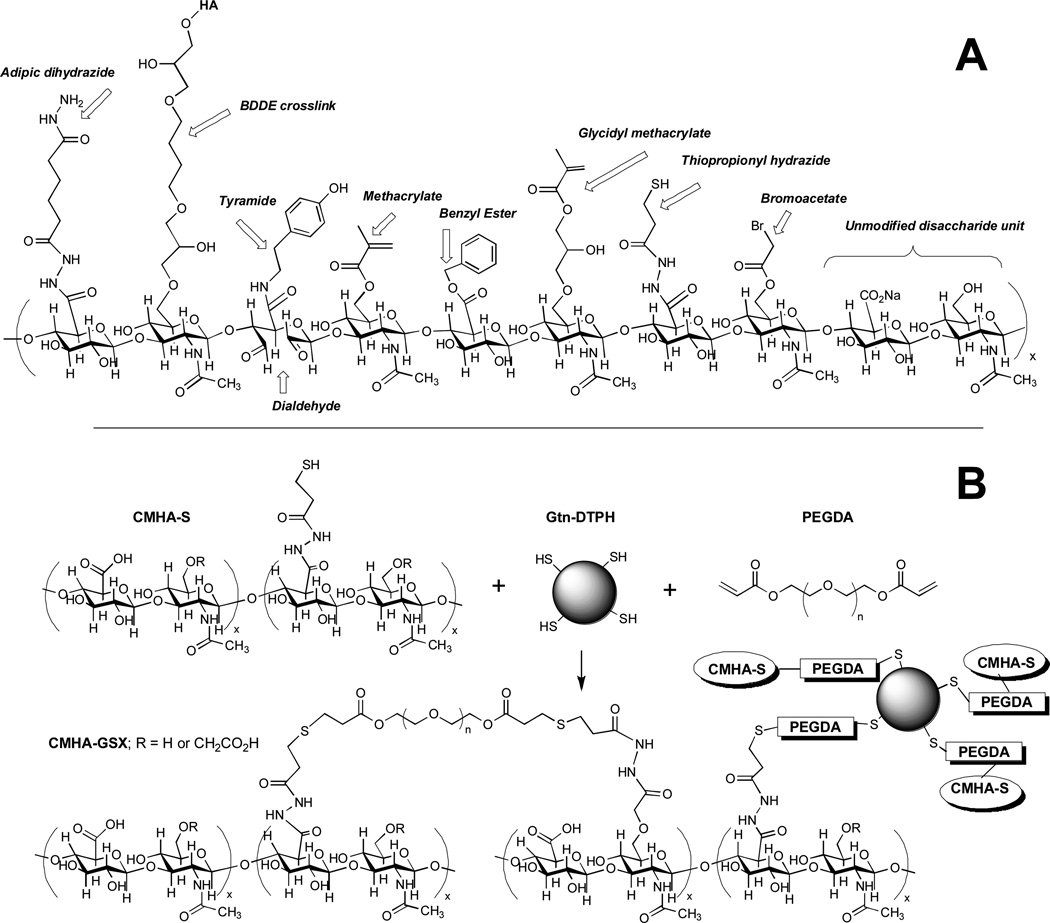
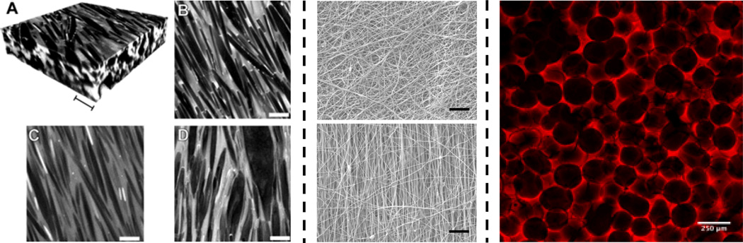
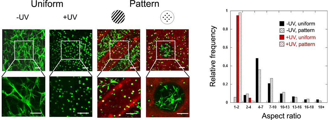
Hyaluronic acid (HA), an immunoneutral polysaccharide that is ubiquitous in the human body, is crucial for many cellular and tissue functions and has been in clinical use for over thirty years. When chemically modified, HA can be transformed into many physical forms-viscoelastic solutions, soft or stiff hydrogels, electrospun fibers, non-woven meshes, macroporous and fibrillar sponges, flexible sheets, and nanoparticulate fluids-for use in a range of preclinical and clinical settings. Many of these forms are derived from the chemical crosslinking of pendant reactive groups by addition/condensation chemistry or by radical polymerization. Clinical products for cell therapy and regenerative medicine require crosslinking chemistry that is compatible with the encapsulation of cells and injection into tissues. Moreover, an injectable clinical biomaterial must meet marketing, regulatory, and financial constraints to provide affordable products that can be approved, deployed to the clinic, and used by physicians. Many HA-derived hydrogels meet these criteria, and can deliver cells and therapeutic agents for tissue repair and regeneration. This progress report covers both basic concepts and recent advances in the development of HA-based hydrogels for biomedical applications.


Figures

Figure 1
Figure 1. Chemical modifications of HA
(A) A hypothetical composite structure illustrating selected primary modifications discussed herein: adipic dihydrazide for use in further crosslinking via acrylamide or hydrazone linkages; butane-1,4-diol diglycidyl ether, a prototypical monolithic crosslinker for HA; tyramide for peroxidase crosslinking; dialdehyde obtained by periodate oxidation; methacrylate on primary 6-hydroxyl group; benzyl ester; glycidyl methacrylate; thiopropionyl hydrazide from DTPH modification; bromoacetate; an unmodified disaccharide unit for comparison. (B) A thioether crosslinked semi-synthetic ECM formed by crosslinking thiol-modified carboxymethyl HA (CMHA-S) with thiol-modified gelatin using the bifunctional crosslinker, PEGDA.
Figure 2
Figure 2. Double-crosslinked HA hydrogels
Hydrogels formed from the crosslinking of particles in a secondary network leading to hierarchical networks with unique microstructures. Reprinted with permission from [26].
Figure 3
Figure 3. Repair of stroke infarct by HA hydrogel-encapsulated NPCs
HyStem-HP gel significantly increased survival and proliferation of murine GFP-tagged embryonic cortex-derived neural progenitor cells (NPCs) injected into the infarct cavity after a photochemically-induced stroke in mouse brain [33]. Of 100,000 injected NPCs, 4000 survived in buffer while 8000 survived in HyStem-HP (p = 0.035). New figure provided by Drs. J. Zhong and S. T. Carmichael.
Figure 4
Figure 4. Delivery of therapeutic antibody-releasing MSCs reduces tumors
Co-encapsulation of wild-type MSCs and HCT-116 colon cancer cells in Extracel-X resulted in robust tumor growth (top). In contrast, use of diabody-releasing MSCs with HCT-116 dramatically suppressed tumor growth. Reprinted with permission from [32].
Figure 5
Figure 5. Mechanical sensitivity of cells to HA hydrogels
Endothelial progenitor cells cultured on HA gels at a range of mechanical properties and two concentrations of VEGF. Capillary-like structures only formed on gels at the higher VEGF concentration and the morphology (e.g., tube length, area, and thickness) was dependent on the gel mechanical properties. Reprinted with permission from [39].
Figure 6
Figure 6. Centrifugal casting of cells in hollow hydrogel cylinders
(Top) The inner walls of a capillary tube or dacron vascular prosthesis are precoated with the Extracel sECM by axial rotation at 2000 rpm (11.2 × g) for 10 min to effect uniform coating during crosslinking and gelation. Then, cells are entrapped between two sECM layers by repeating the process with a cell suspension, giving a concentric sandwich construct. Panels b shows a gel-coated dacron vascular graft, and panel c shows GFP-labeled QCE-6 quail vascular progenitor cells. Partially reprinted with permission from [57].
Figure 7
Figure 7. Dynamic crosslinking of macromolecular thiols with gold nanoparticles
(A) Au-NPs act as multivalent crosslinkers for thiol-modified HA. (B) Bioprinting consists of deposition of acellular AuNP-CMHA-S gels (blue) and cell-containing AuNP-CMHA-S/Gelatin-DTPH gels to produce a cylindrical structure. (C) A tubular cellularized construct printed without the central core outer annulus of the acellular gel. Partially reprinted with permission from [66].
Figure 8
Figure 8. Structures of a range of photopolymerizable HA macromers
HA macromers can be synthesized to include reactive methacrylate groups either directly (MeHA) or with a hydrolytically degradable spacer of lactic acid (MeLAHA) or caprolactone (MeCLHA). When photocrosslinked, these macromers form hydrogels with varied degradation behavior (measured with release of uronic acid), with degradation rates of MeLAHA > MeCLHA > MeHA.
Figure 9
Figure 9. Examples of photocrosslinked HA scaffold structures
Photopolymerizable HA macromers can be processed into a range of structures based on: crystal templating (left, scale bars = 10 µm), electrospinning fibrous structures (middle, non-aligned on top, aligned on bottom, scale bar = 10 µm) and macroporous scaffolds from sphere templating (right, scale bar = 250 µm). For crystal templating, confocal images (A: reconstruction, B–C: scan) of hydrogels containing urea crystals before (A, B) and after (C: swollen, D: dry) crystal removal are shown. Partially reprinted with permission from [128].
Figure 10
Figure 10. Spatially controlled behavior of stem cells in 3D hydrogels
Human mesenchymal stem cells (confocal images on left, quantification of aspect ratios on right) were encapsulated in HA hydrogels using MMP-cleavable crosslinkers using a sequential crosslinking process. The introduction of light introduces kinetic chains in a spatially controlled manner (illustrated in red) that alters the ability of a cell to remodel the hydrogel, leading to spatially controlled cell spreading. Reprinted with permission from [134].

Similar articles

Cited by

References

    1. Fraser JR, Laurent TC, Laurent UB. J Intern Med. 1997;242:27. - PubMed
    1. Toole BP. Semin Cell Dev Biol. 2001;12:79. - PubMed
    1. Toole BP. Nat Rev Cancer. 2004;4:528. - PubMed
    1. Laurent TC, Fraser JR. Ciba Found Symp. 1986;124:9. - PubMed
    1. Kuo JW. Practical aspects of hyaluronan based medical products. Boca Raton: CRC/Taylor & Francis; 2006.

Publication types

LinkOut - more resources