

Systematic review of the concurrent and predictive validity of MRI biomarkers in OA
- PMID: 21396463
- PMCID: PMC3268360
- DOI: 10.1016/j.joca.2010.10.029
Systematic review of the concurrent and predictive validity of MRI biomarkers in OA
Abstract
Objective: To summarize literature on the concurrent and predictive validity of MRI-based measures of osteoarthritis (OA) structural change.
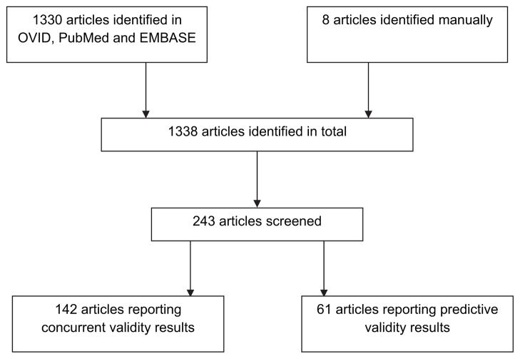
Methods: An online literature search was conducted of the OVID, EMBASE, CINAHL, PsychInfo and Cochrane databases of articles published up to the time of the search, April 2009. 1338 abstracts obtained with this search were preliminarily screened for relevance by two reviewers. Of these, 243 were selected for data extraction for this analysis on validity as well as separate reviews on discriminate validity and diagnostic performance. Of these 142 manuscripts included data pertinent to concurrent validity and 61 manuscripts for the predictive validity review. For this analysis we extracted data on criterion (concurrent and predictive) validity from both longitudinal and cross-sectional studies for all synovial joint tissues as it relates to MRI measurement in OA.
Results: Concurrent validity of MRI in OA has been examined compared to symptoms, radiography, histology/pathology, arthroscopy, CT, and alignment. The relation of bone marrow lesions, synovitis and effusion to pain was moderate to strong. There was a weak or no relation of cartilage morphology or meniscal tears to pain. The relation of cartilage morphology to radiographic OA and radiographic joint space was inconsistent. There was a higher frequency of meniscal tears, synovitis and other features in persons with radiographic OA. The relation of cartilage to other constructs including histology and arthroscopy was stronger. Predictive validity of MRI in OA has been examined for ability to predict total knee replacement (TKR), change in symptoms, radiographic progression as well as MRI progression. Quantitative cartilage volume change and presence of cartilage defects or bone marrow lesions are potential predictors of TKR.
Conclusion: MRI has inherent strengths and unique advantages in its ability to visualize multiple individual tissue pathologies relating to pain and also predict clinical outcome. The complex disease of OA which involves an array of tissue abnormalities is best imaged using this imaging tool.
Copyright © 2011 Osteoarthritis Research Society International. All rights reserved.
Conflict of interest statement
David Hunter receives research or institutional support from DonJoy, NIH, and Stryker.
Other authors declared no conflict of interest
Figures
References
-
- Kellgren JH, Lawrence JS. Atlas of standard radiographs. Oxford: Blackwell Scientific; 1963.
-
- Guermazi A, Burstein D, Conaghan P, Eckstein F, Hellio Le Graverand-Gastineau MP, Keen H, et al. Imaging in osteoarthritis. Rheum Dis Clin North Am. 2008;34:645–87. - PubMed
-
- Dieppe PA, Lohmander LS. Pathogenesis and management of pain in osteoarthritis. Lancet. 2005;365:965–73. [Review] [100 refs] - PubMed
-
- Hannan MT, Felson DT, Pincus T. Analysis of the discordance between radiographic changes and knee pain in osteoarthritis of the knee. J Rheumatol. 2000;27:1513–7. - PubMed
Publication types
MeSH terms
Substances
Grants and funding
LinkOut - more resources
Full Text Sources
Medical
Miscellaneous
