. 2011 Apr 19;20(4):526-39.
doi: 10.1016/j.devcel.2011.02.010. Epub 2011 Mar 10.

Blood vessel tubulogenesis requires Rasip1 regulation of GTPase signaling

Affiliations

Blood vessel tubulogenesis requires Rasip1 regulation of GTPase signaling

Ke Xu et al. Dev Cell. .

Abstract

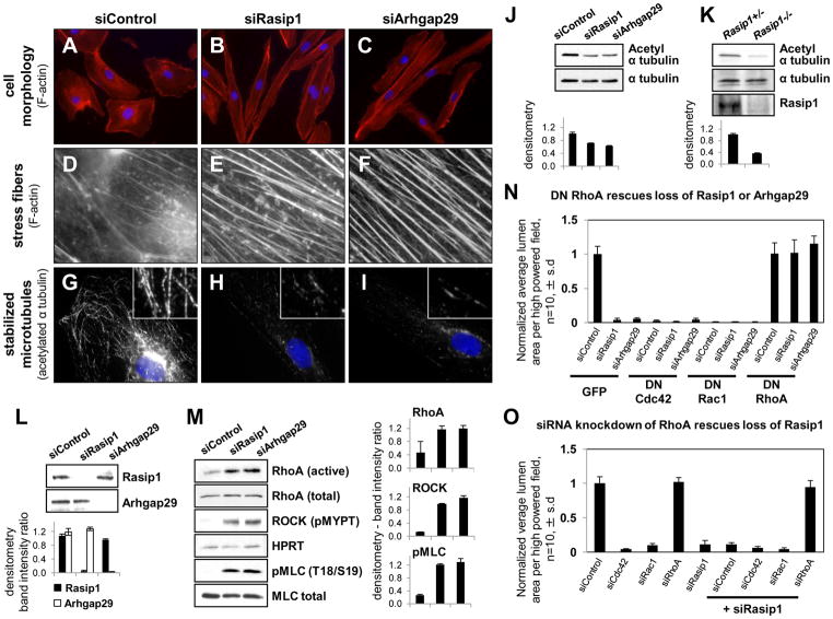
Cardiovascular function depends on patent blood vessel formation by endothelial cells (ECs). However, the mechanisms underlying vascular "tubulogenesis" are only beginning to be unraveled. We show that endothelial tubulogenesis requires the Ras interacting protein 1, Rasip1, and its binding partner, the RhoGAP Arhgap29. Mice lacking Rasip1 fail to form patent lumens in all blood vessels, including the early endocardial tube. Rasipl null angioblasts fail to properly localize the polarity determinant Par3 and display defective cell polarity, resulting in mislocalized junctional complexes and loss of adhesion to extracellular matrix (ECM). Similarly, depletion of either Rasip1 or Arhgap29 in cultured ECs blocks in vitro lumen formation, fundamentally alters the cytoskeleton, and reduces integrin-dependent adhesion to ECM. These defects result from increased RhoA/ROCK/myosin II activity and blockade of Cdc42 and Rac1 signaling. This study identifies Rasip1 as a unique, endothelial-specific regulator of Rho GTPase signaling, which is essential for blood vessel morphogenesis.


Figures

Figure 1
Figure 1. Rasip1 is essential for vascular tubulogenesis in all blood vessels
(A–D) Rasip1 expression is conserved in the embryonic vasculature across species shown by in situ hybridization; mouse (A, E9.5; B, E8.5 transverse section, left dorsal aorta), Xenopus tropicalis (C, st.30), and zebrafish (D, 24hpf). (E–H) E9.5 littermate mouse embryos showing defects in Rasip1/ embryos (F) and yolk sacs (H). (I–L) Whole mount Flk1-lacZ beta-galactosidase staining of Rasip1+/ (I, K) and Rasip1/ (J, L) yolk sacs (I, J) and embryonic heads (K, L), showing narrow Rasip1/ blood vessels that fail to remodel. (M–P) Connexin40 (Cx40 or Gja5) (M, N) or EphB4-lacZ (O, P) in littermate E9.5 Rasip1+/ and Rasip1/ yolk sacs (M, N) or embryos (O, P) showing failed arterial (M, N) and venous (O, P) differentiation in Rasip1/ vessels. (Q–Z′) Flk1-LacZ beta-galactosidase staining of littermate E8.5 (6 somite stage) Rasip1+/ and Rasip1/ embryos and tissues, in whole mount or section. (Q, R) Ventral views of E8.5 embryonic paired dorsal aortae. Anterior is up. (S–V) Transverse sections of E8.5 embryos, through the trunk region, posterior to the heart. (U, V) Closeup views, showing open lumens in Rasip1+/;Flk1-lacZ embryos (S, U), but lumenless cords in Rasip1/;Flk1-lacZ embryos (T, V). Sections through hearts of E8.5 (W, X), showing absence of a lumen in the endocardium of Rasip1/;Flk1-lacZ embryos (X). Yolk sacs in whole mount (Y, Z) or section views (Y′, Z′). Vascular tubes are absent in all Rasip1/ vessels examined. Arrows, dorsal aortae. Arrowheads, yolk sac vessels. ys: yolk sac. Scale bars: 500μm (A, E–H), 250μm (C), 200μm (K–P), 100μm (D, I, J, Q, R, Y, Z), 50μm (B, S–Z, Y′, Z′). See also Figures S1 and S2.
Figure 2
Figure 2. Rasip1 and Arhgap29 are required for in vitro EC lumen formation and regulation of small GTPase signaling
(A) Silver staining of the affinity-purified Rasip1 complex. Marked bands were identified by mass spectrometry. (B) Arhgap29 protein in E8.5 mouse embryonic aortic ECs, ventral view. Arrows, paired dorsal aortae. Scale bar: 50μm. (C) Rasip1 and Arhgap29 expression in cultured EC (MS1). Arrowheads, Rasip1/Arhgap29 co-localization in punctae. Nuclei, DAPI (blue). (D) Western blots of Rasip1 and Arhgap29 following siRNA treatment of cultured HUVECs in 3D collagen matrices. (E) Lumen formation is blocked in siRasip1-/siArhgap29-treated HUVECs cultured in 3D collagen matrices. Arrowheads, visible EC lumen. (F) Quantification of lumen formation in control- or siRasip1-/siAhgap29- treated HUVECs. (G) Cdc42, Rac1 and RhoA activities and (H) reduction of kinase signaling downstream of Cdc42/Rac1 pathway in siRNA-treated HUVECs in 3D cultures at 24hrs. Error bars represent standard deviation. See also Figure S3.
Figure 3
Figure 3. Rasip1 and Arhgap29 regulate EC architecture and tubulogenesis through Rho family GTPases
(A–F) Immunofluorescent phalloidin staining showing gross cell morphology and stress fibers in siRNA-treated HUVECs. (G–I) ‘Stabilized’ microtubule shown by acetylated α-tubulin staining in siRNA-treated HUVECs. Insets, high magnification view of microtubules. Nuclei, DAPI (blue). (J, K) Acetylated α–tubulin level is down regulated in siRNA treated ECs in vitro (J) and in vivo in E9.5 Rasip1/ embryos (K), n = 2. (L, M) Western blots showing RhoA and ROCK activity (pMYPT, T696), phosphorylation of myosin light chain (pMLC, T18/S19) in siRNA-treated HUVECs, n = 3. (N) Dominant negative (DN) RhoA rescues in vitro lumenless phenotype in 3D collagen matrices in siRasip1-/siArhgap29-treated HUVECs. (O) Loss of Rasip1, which causes a lumenless EC phenotype in 3D collagen matrices, is rescued by siRNA knockdown of RhoA. Error bars represent standard deviation. See also Figure S4.
Figure 4
Figure 4. Rasip1 and Arhgap29 are required for maturation of endothelial-ECM adhesion
(A–F) Immature focal adhesions shown by phosphorylated Paxillin (Y118) staining in siRNA-treated HUVECs. (G) Phosphorylated Paxillin (Y118) is up regulated in siRNA treated ECs in vitro and in vivo in Rasip1/ embryos, n = 2. (H) Flow cytometry showing decrease in activated β1 integrin (9EG7) in the absence of Rasip1 or Arhgap29. MnCl2 (5mM) treatment reveals total surface levels of integrins are unchanged. (I–K) Mature fibrillar adhesions shown by activated β1 integrin (9EG7) expression in siRNA-treated HUVECs. Insets, high magnification view of 9EG7+ FBs. Nuclei, DAPI (blue). Error bars represent standard deviation. See also Figures S5 and S6.
Figure 5
Figure 5. Rasip1−/− angioblasts remain cuboidal and fail to adhere to surrounding ECM
(A–N) Confocal immunofluorescence microscopy of 36 somite Rasip1+/− (A, C, E, G, I, K, M) and Rasip1−/− (B, D, F, H, J, L, N) dorsal aortae (Flk1-EGFP, A, B, G, H, IN) in transverse sections: (A, B) claudin-5, (C, D) ZO-1 and laminin, (E, F) VE-cadherin and collagen IV, and (G, H) fibronectin. Nuclei, DAPI (blue). (O) Average dorsal aortae diameter at 68 somite stage, n=30. Arrows, EC-EC junctions in Rasip1+/ embryos. Arrowheads, mislocalized EC-EC junctions. Scale bars, 25 μm. Error bars represent standard deviation.
Figure 6
Figure 6. Rasip1−/− angioblasts display defective cell polarity and fail to localize junctional proteins to cord periphery
(A–D, G–L) Confocal immunofluorescence microscopy of Rasip1+/− (A, C, G, I, K) and Rasip1−/− (B, D, H, J, L) aortic ECs in transverse sections: podocalyxin (green), ZO-1 (red), and Par3 (G–L, red). Nuclei, DAPI (blue). (E, F) Rasip1 null aortic cord angioblasts display mislocalized EC-EC TJs. TEM images of transverse sections of Rasip1+/− (E) and Rasip1−/− (F) littermates, at 4-somite stage. (E, F) Black arrows, tight junctions; white arrows, slits/isolated lumens between ECs. (A″, D″, J) Arrows, ectopic basal TJs; arrowheads, ectopic apical TJs; dotted line, basal endoderm surface. Scale bars, 5 μm (A–J), 20μm(K, L). See also Figure S7.
Figure 7
Figure 7. Models of Rasip1 regulation of embryonic vascular tubulogenesis
(A) Rasip1 regulates EC contractility and adhesion by modulating Rho family small GTPases. (B, C) The balance between EC-EC and EC-ECM adhesion is critical to vascular cord to tube transition. (B) Loss of integrin adhesion at the basal cell surface and (C) failure of junctional protein redistribution from the apical cell surface to the cord periphery result in failed vascular tubulogenesis.

Comment in

  • LUMENating blood vessels.
    Iruela-Arispe ML. Iruela-Arispe ML. Dev Cell. 2011 Apr 19;20(4):412-4. doi: 10.1016/j.devcel.2011.03.020. Dev Cell. 2011. PMID: 21497753 Free PMC article.

References

    1. Aird WC. Phenotypic heterogeneity of the endothelium: II. Representative vascular beds. Circ Res. 2007;100:174–190. - PubMed
    1. Andrew DJ, Ewald AJ. Morphogenesis of epithelial tubes: Insights into tube formation, elongation, and elaboration. Dev Biol. 341:34–55. - PMC - PubMed
    1. Bayless KJ, Davis GE. The Cdc42 and Rac1 GTPases are required for capillary lumen formation in three-dimensional extracellular matrices. J Cell Sci. 2002;115:1123–1136. - PubMed
    1. Bayless KJ, Davis GE. Microtubule depolymerization rapidly collapses capillary tube networks in vitro and angiogenic vessels in vivo through the small GTPase Rho. J Biol Chem. 2004;279:11686–11695. - PubMed
    1. Bazzoni G, Shih DT, Buck CA, Hemler ME. Monoclonal antibody 9EG7 defines a novel beta 1 integrin epitope induced by soluble ligand and manganese, but inhibited by calcium. J Biol Chem. 1995;270:25570–25577. - PubMed

Publication types