. 2011 Jul;23(7):1170-8.
doi: 10.1016/j.cellsig.2011.03.004. Epub 2011 Mar 17.

Nuclear localization of pyruvate dehydrogenase complex-E2 (PDC-E2), a mitochondrial enzyme, and its role in signal transducer and activator of transcription 5 (STAT5)-dependent gene transcription

Affiliations

Nuclear localization of pyruvate dehydrogenase complex-E2 (PDC-E2), a mitochondrial enzyme, and its role in signal transducer and activator of transcription 5 (STAT5)-dependent gene transcription

Fu-Yu Chueh et al. Cell Signal. 2011 Jul.

Abstract

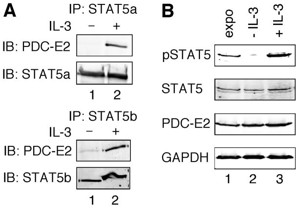
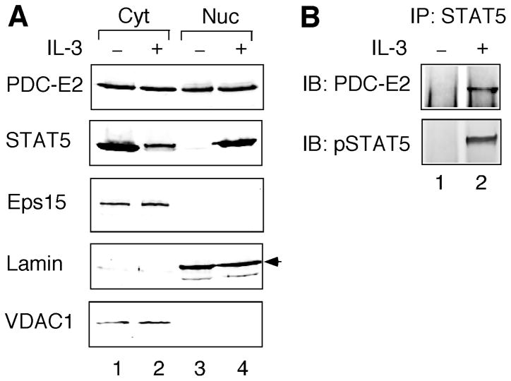
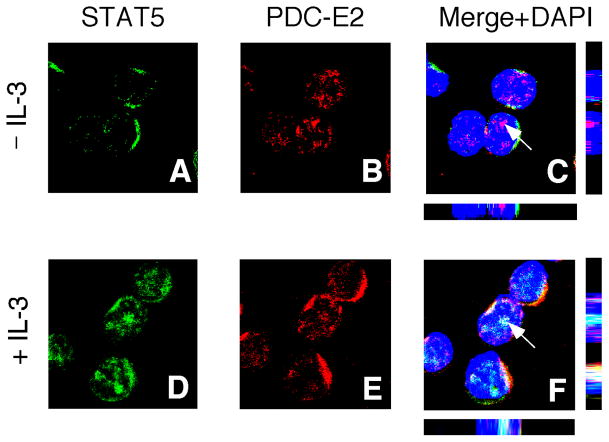
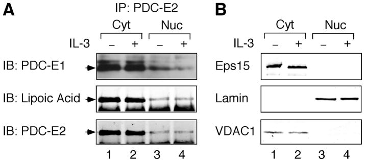
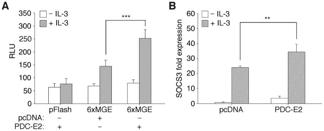
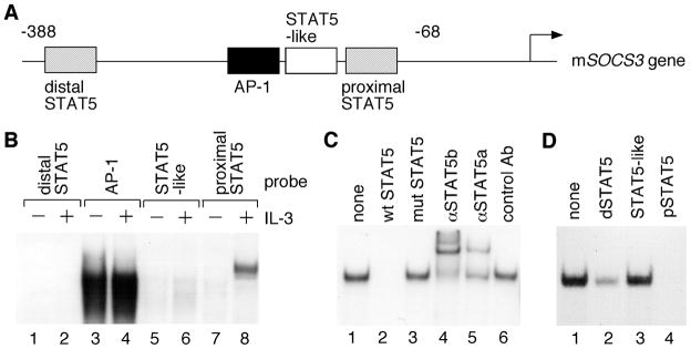
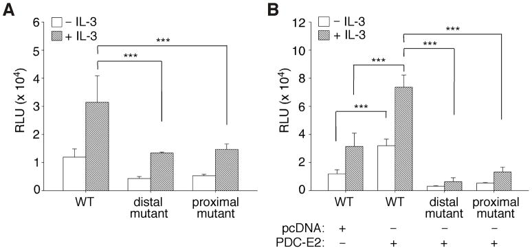
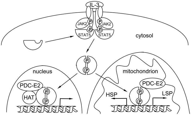
STAT (signal transducer and activator of transcription) proteins play a critical role in cellular response to a wide variety of cytokines and growth factors by regulating specific nuclear genes. STAT-dependent gene transcription can be finely tuned through the association with co-factors in the nucleus. We showed previously that STAT5 (including 5a and 5b) specifically interacts with a mitochondrial enzyme PDC-E2 (E2 subunit of pyruvate dehydrogenase complex) in both leukemic T cells and cytokine-stimulated cells. However, the functional significance of this novel association remains largely unknown. Here we report that PDC-E2 may function as a co-activator in STAT5-dependent nuclear gene expression. Subcellular fractionation analysis revealed that a substantial amount of PDC-E2 was constitutively present in the nucleus of BaF3, an interleukin-3 (IL-3)-dependent cell line. IL-3-induced tyrosine-phosphorylated STAT5 associated with nuclear PDC-E2 in co-immunoprecipitation analysis. These findings were confirmed by confocal immunofluorescence microscopy showing constant nuclear localization of PDC-E2 and its co-localization with STAT5 after IL-3 stimulation. Similar to mitochondrial PDC-E2, nuclear PDC-E2 was lipoylated and associated with PDC-E1. Overexpression of PDC-E2 in BaF3 cells augmented IL-3-induced STAT5 activity as measured by reporter assay with consensus STAT5-binding sites. Consistent with the reporter data, PDC-E2 overexpression in BaF3 cells led to elevated mRNA levels of endogenous SOCS3 (suppressor of cytokine signaling 3) gene, a known STAT5 target. We further identified two functional STAT5-binding sites in the SOCS3 gene promoter important for its IL-3-inducibility. The observation that both cis-acting elements were essential to detect the stimulatory effect by PDC-E2 strongly supports the role of PDC-E2 in up-regulating the transactivating ability of STAT5. All together, our results reveal a novel function of PDC-E2 in the nucleus. It also raises the possibility of nuclear-mitochondrial crosstalk through the interaction between STAT5 and PDC-E2.


Figures

Fig. 1
Fig. 1. PDC-E2 binds to STAT5a and STAT5b in IL-3-stimulated BaF3 cells
(A) Whole cell lysates were prepared from IL-3-deprived BaF3 cells either left untreated or stimulated with IL-3 for 15 min. Equal amounts of total proteins were immunoprecipitated (IP) with antibody specific for STAT5a (upper panels) or STAT5b (lower panels). Eluted proteins were resolved by SDS-PAGE and subjected to immunoblotting (IB) using anti-PDC-E2 antibody. Membranes were then stripped and reblotted with antibodies specific for STAT5a or STAT5b to confirm equal amounts of STAT5 proteins in the immunoprecipitates. (B) Whole cell lysates were prepared from exponentially growing (expo), IL-3-deprived (− IL-3), or IL-3-stimulated (+ IL-3) BaF3 cells. Equal amounts of total proteins were subjected to immunoblotting using antibodies specific for tyrosine-phosphorylated STAT5 (pSTAT5), STAT5, PDC-E2 and GAPDH.
Fig. 2
Fig. 2. PDC-E2 localizes in the nucleus and interacts with tyrosine-phosphorylated STAT5 in IL-3-stimulated BaF3 cells
BaF3 cells without or with 15 min of IL-3 stimulation were subjected to subcellular fractionation to separate the nuclei (Nuc) from cytosol (Cyt). (A) Normalized total proteins from both fractions were resolved by SDS-PAGE, followed by Western blot analysis with antibodies specific for PDC-E2, STAT5, Eps15, Lamin, and VDAC1. The arrowhead on the right indicates the correct position of Lamin. (B) Normalized nuclear proteins were immunoprecipitated with anti-STAT5 antibody, followed by immunoblotting with antibodies specific for PDC-E2 and tyrosine-phosphorylated STAT5 (pSTAT5).
Fig. 3
Fig. 3. Subcellular localization of PDC-E2 and its co-localization with STAT5 in BaF3 cells
IL-3-deprived BaF3 cells were either left unstimulated (panels A–C) or stimulated with IL-3 for 15 min (panels D–F). Fixed and permeabilized cells were stained with anti-PDC-E2 polyclonal antibody and anti-STAT5 monoclonal antibody, followed by the corresponding secondary antibodies conjugated with different fluorophores (green for STAT5 and red for PDC-E2). Nuclei were visualized using DAPI (blue) as a counterstain. The three-color merge images are shown in panels C and F. The arrowheads denote the points of confocal analysis through the z axes with the bottom and side bars representing the xz and yz axes, respectively. Yellow color indicates co-localization of PDC-E2 and STAT5 outside nuclei. White color shows co-localization of PDC-E2 and STAT5 within nuclei.
Fig. 4
Fig. 4. Nuclear PDC-E2 is lipoylated and binds to PDC-E1 constitutively in BaF3 cells
Cytosolic and nuclear fractions were prepared from IL-3-deprived and IL-3-stimulated BaF3 cells as described for Fig. 2. (A) Total proteins were immunoprecipitated with anti-PDC-E2 antibody followed by immunoblotting using antibodies specific for PDC-E1, lipoic acid, and PDC-E2. PDC-E1 immunoblotting was performed using Luminata Forte high-sensitivity chemiluminescent system. The arrowheads on the left indicate the correct positions of target proteins. (B) Normalized proteins from cytosolic and nuclear fractions were analyzed by immunoblotting using antibodies specific for Eps15, Lamin, and VDAC1.
Fig. 5
Fig. 5. IL-3-induced STAT5 reporter activity and endogenous SOCS3 expression is augmented by exogenous PDC-E2
(A) BaF3 cells were electroporated with firefly luciferase reporter constructs without (pFlash) or with six consecutive mammary gland elements (6xMGE) and co-transfected with either pcDNA vector control or PDC-E2 expression construct. Dual luciferase assay was conducted in transfected cells before and after 16 h of IL-3 stimulation. Data were normalized to renilla luciferase control and expressed as relative luciferase units (RLU). (B) BaF3 cells were transfected with pcDNA vector control or PDC-E2 expression construct as described above. Total RNAs were isolated from transfected cells before and after 30 min of IL-3 stimulation, reverse transcribed, and then subjected to real time PCR using primers specific for mouse SOCS3 and actin mRNAs. Data were normalized to actin and expressed as fold change to vector-transfected cells before IL-3 stimulation. ** P < 0.01, *** P < 0.001.
Fig. 6
Fig. 6. Characterization of the mouse SOCS3 promoter
(A) Schematic diagram of four potential cis-regulatory elements in the mouse SOCS3 promoter. (B) Nuclear extracts were prepared from IL-3-deprived BaF3 cells either left untreated or stimulated with IL-3 for 30 min. Equal amounts of nuclear proteins were subjected to EMSA with 32P-labeled oligonucleotides containing the four cis-acting elements as described in A. (C) Nuclear extracts from IL-3-stimulated BaF3 cells were incubated with a 32P-labeled probe containing the proximal STAT5 site (lane 1). Competition assays were conducted in the presence of 100-fold molar excess of unlabeled oligonucleotides that contain either the wild-type (lane 2) or the mutated proximal STAT5 site (lane 3). Supershift assays were performed with control antibody or antibodies specific for STAT5a and STAT5b (lanes 4–6). (D) Nuclear extracts from IL-3-stimulated BaF3 cells were incubated with a 32P-labeled consensus STAT5 probe derived from the sheep β-casein gene promoter (lane 1). Competition assays were carried out with 100-fold molar excess of unlabeled oligonucleotides derived from the distal STAT5 site (lane 2), the STAT5-like site (lane 3) or the proximal STAT5 site (lane 4) in the mouse SOCS3 gene promoter.
Fig. 7
Fig. 7. Combined effects of IL-3 and PDC-E2 on the SOCS3 promoter are mediated through both proximal and distal STAT5 sites
Dual-luciferase reporter assays were conducted in IL-3-deprived and IL-3-stimulated BaF3 cells as described for Fig. 5A. (A) BaF3 cells were transfected with firefly luciferase reporter driven by the wild-type mSOCS3 promoter (WT), the promoter with mutated distal STAT5 site, or the promoter with mutated proximal STAT5 site. (B) BaF3 cells were co-transfected with PDC-E2 expression construct or pcDNA vector control in addition to the reporter plasmids as described in A. *** P < 0.001.
Fig. 8
Fig. 8. A hypothetical model of nuclear-mitochondrial crosstalk through cytokine-induced STAT5 and PDC-E2 interaction
Upon IL-3 stimulation, receptor dimerization induces tyrosine phosphorylation of the associated JAK2 tyrosine kinase. Active JAK2 phosphorylates the cytoplasmic tails of receptor subunits. Cytoplasmic STAT5 is recruited to the receptor complex and phosphorylated by the JAK2 kinase. Tyrosine-phosphorylated STAT5 proteins dimerize and translocate to the nucleus and the mitochondrion. In the nucleus, tyrosine-phosphorylated STAT5 binds to promoter regions of distinct target genes, such as SOCS3. The associated PDC-E2 may work in concert with histone acetyltransferase (HAT) to enhance STAT5-dependent nuclear gene expression. On the other hand, binding of tyrosine-phosphorylated STAT5 to the control region of mitochondrial DNA may modulate transcription initiated from the heavy strand promoter (HSP) and light strand promoter (LSP). Phosphorylation of key tyrosine residues are represented by “P” in circles.

References

    1. Levy DE, Darnell JE., Jr Nat Rev Mol Cell Biol. 2002;3:651–662. - PubMed
    1. Schindler C, Levy DE, Decker T. J Biol Chem. 2007;282:20059–20063. - PubMed
    1. Lanning NJ, Carter-Su C. Rev Endocr Metab Dis. 2006;7:225–235. - PubMed
    1. Yu H, Jove R. Nat Rev Cancer. 2004;4:97–105. - PubMed
    1. Shuai K. Oncogene. 2000;19:2638–2644. - PubMed

Publication types

MeSH terms

Substances

LinkOut - more resources