Comparative Study
. 2011 Sep 30;178(3):458-64.
doi: 10.1016/j.resp.2011.03.003. Epub 2011 Mar 17.

Vagal afferents modulate cytokine-mediated respiratory control at the neonatal medulla oblongata

Affiliations
Comparative Study

Vagal afferents modulate cytokine-mediated respiratory control at the neonatal medulla oblongata

Kannan V Balan et al. Respir Physiol Neurobiol. .

Abstract

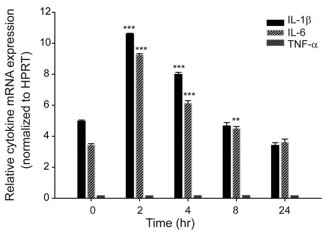
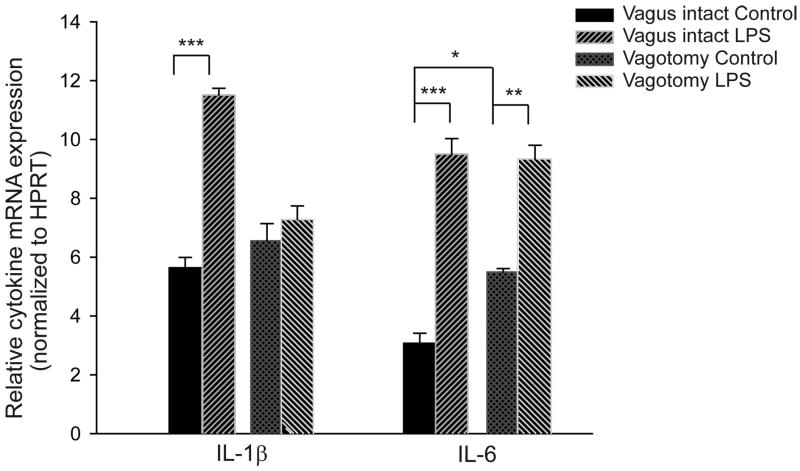
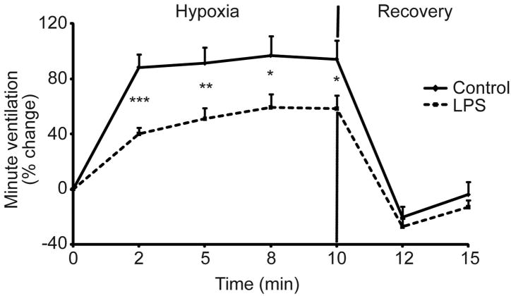
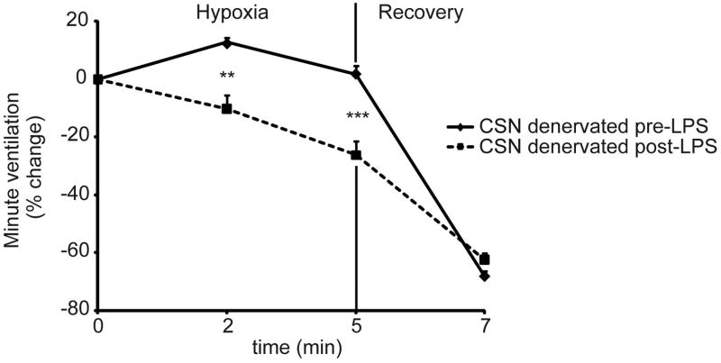
Perinatal sepsis and inflammation trigger lung and brain injury in preterm infants, and associated apnea of prematurity. We hypothesized that endotoxin exposure in the immature lung would upregulate proinflammatory cytokine mRNA expression in the medulla oblongata and be associated with impaired respiratory control. Lipopolysaccharide (LPS, 0.1mg/kg) or saline was administered intratracheally to rat pups and medulla oblongatas were harvested for quantifying expression of mRNA for proinflammatory cytokines. LPS-exposure significantly increased medullary mRNA for IL-1β and IL-6, and vagotomy blunted this increase in IL-1β, but not IL-6. Whole-body flow plethysmography revealed that LPS-exposed pups had an attenuated ventilatory response to hypoxia both before and after carotid sinus nerve transection. Immunochemical expression of IL-1β within the nucleus of the solitary tract and area postrema was increased after LPS-exposure. In summary, intratracheal endotoxin-exposure in rat pups is associated with upregulation of proinflammatory cytokines in the medulla oblongata that is vagally mediated for IL-1β and associated with an impaired hypoxic ventilatory response.


Figures

Figure 1
Figure 1
Time course for IL-1β, IL-6 and TNF-α mRNA expression in the medulla oblongata following intratracheal instillation of LPS (0.1mg/kg). Samples were collected 2, 4, 8 and 24 hr post instillation. Note, the 2 hr time point showed optimal induction for IL-1β and IL-6 mRNA, whereas no quantifiable expression for TNF-α at our dose of LPS was observed in this experimental model. Proinflammatory cytokine expressions were normalized to hypoxanthine phophoribosyltransferase (HPRT), a constitutively expressed housekeeping gene. Data are expressed as mean ± S.E.M, n = 6 per group. ** = p<0.01; *** = p<0.001 compared to baseline at time zero.
Figure 2
Figure 2
Relative IL-1β and IL-6 mRNA expression in the medulla oblongata two hours after saline or LPS instillation in vagus intact or vagotomized pups. LPS instillation significantly increased IL-1β mRNA expression in the medulla oblongata which was not apparent after vagotomy. In contrast vagotomy did not appear to diminish the increase in IL-6 mRNA expression after LPS treatment. Vagotomy per se did not alter the level of baseline medullary IL-1β mRNA expression but did increase baseline IL-6 expression. Proinflammatory cytokine expressions were normalized to the expression of HPRT, a constitutively expressed housekeeping gene. Data are expressed as mean ± S.E.M, n = 10 per group. * = p<0.05; ** = p<0.01; *** = p<0.001
Figure 3
Figure 3
Effect of intratracheal LPS on minute ventilation during exposure to 10% O2 in 10– 12 day-old F344 rats (n = 10 per group). Percentage change in minute ventilation from baseline was recorded in room air (time zero), in response to 10% O2 exposure, and during post-hypoxic response in room air using whole body plethysmography. Note, the hypoxic ventilatory response was significantly decreased in LPS as compared to saline instilled rat pups. Data are expressed as mean ± S.E.M. * = p<0.05, ** = p<0.01 and *** = p<0.001 between groups for the same time point.
Figure 4
Figure 4
Effect of LPS on minute ventilation during exposure to 5% CO2 in 10 – 12 day-old F344 rats (n = 10 per group). Percentage change in minute ventilation from baseline was recorded in room air (time zero), in response to 5% CO2 exposure, and during post-hypercapnic response in room air using whole body plethysmography. Data are expressed as mean ± S.E.M. Note, no significant difference in hypercapnia ventilatory response was observed between saline and LPS instilled rat pups.
Figure 5
Figure 5
Hypoxia-induced ventilatory changes in carotid sinus nerve (CSN)-transected pups prior to (continuous line) and 2 – 3 hr post LPS treatment (interrupted line) (n = 6 per group). Minute ventilation (represented as percentage change from the baseline) was recorded during 10% O2 exposure followed by recovery in room air using whole body flow plethysmography. LPS provoked a significant ventilatory depressant response to hypoxia in CSN-denervated rat pups. Data are expressed as mean ± S.E.M. ** = p<0.01 and *** = p<0.001 between groups for the same time point.
Figure 6
Figure 6
A: Schematic diagram showing regions of localized IL-1β in the medulla oblongata [area postrema (AP); nucleus tractus solitarius (nTS)]. B: Photomicrographs showing localization of IL-1β two to three hr following tracheal instillation of saline (control) or LPS in the AP (top row) and nTS (bottom row). Arrows represent IL-1β immunostained cells and a higher magnification for IL-1β immunoreactivity in the AP and nTS is presented for LPS-instilled pups. Note, LPS increased IL-1β expression in both the AP and nTS. Scale bar represents 50 μm.

References

    1. Abu-Shaweesh JM, Martin RJ. Neonatal apnea: what’s new? Pediatr Pulmonol. 2008;43:937–944. - PubMed
    1. Brady LS, Lynn AB, Herkenham M, Gottesfeld Z. Systemic interleukin-1 induces early and late patterns of c-fos mRNA expression in brain. J Neurosci. 1994;14:4951–4964. - PMC - PubMed
    1. Brattsand R, Linden M. Cytokine modulation by glucocorticoids: mechanisms and actions in cellular studies. Aliment Pharmacol Ther. 1996;10(Suppl 2):81–90. discussion 91–82. - PubMed
    1. Chai Z, Gatti S, Toniatti C, Poli V, Bartfai T. Interleukin (IL)-6 gene expression in the central nervous system is necessary for fever response to lipopolysaccharide or IL-1 beta: a study on IL-6-deficient mice. J Exp Med. 1996;183:311–316. - PMC - PubMed
    1. Chan RK, Brown ER, Ericsson A, Kovacs KJ, Sawchenko PE. A comparison of two immediate-early genes, c-fos and NGFI-B, as markers for functional activation in stress-related neuroendocrine circuitry. J Neurosci. 1993;13:5126–5138. - PMC - PubMed

Publication types