. 2011 Mar 11;88(3):351-61.
doi: 10.1016/j.ajhg.2011.02.007.

A recurrent deletion of DPY19L2 causes infertility in man by blocking sperm head elongation and acrosome formation

Affiliations

A recurrent deletion of DPY19L2 causes infertility in man by blocking sperm head elongation and acrosome formation

Radu Harbuz et al. Am J Hum Genet. .

Abstract

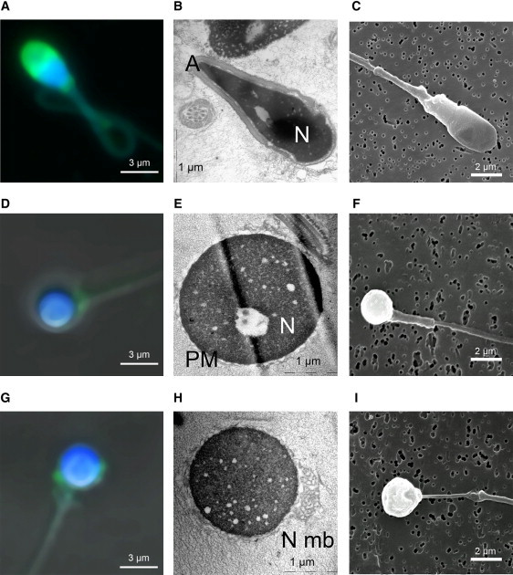
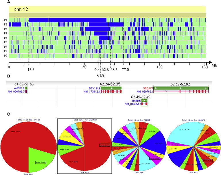
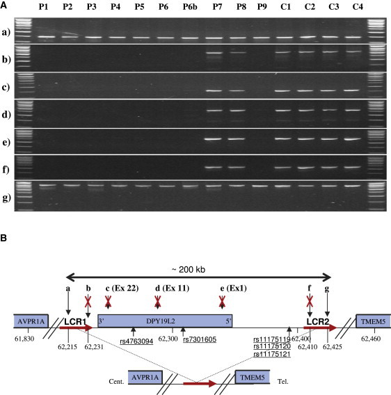
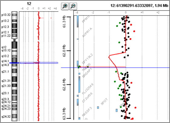
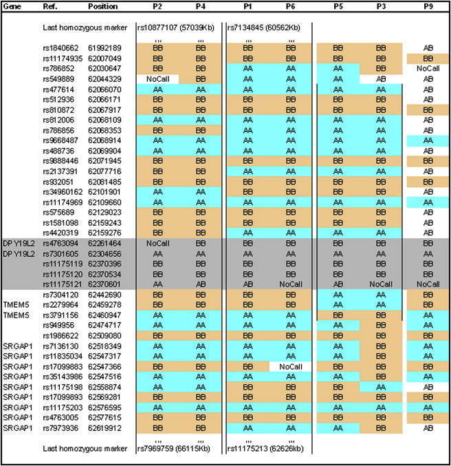
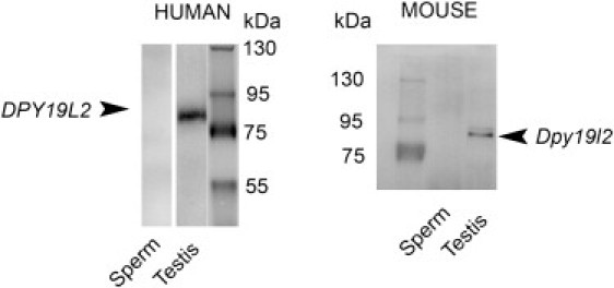
An increasing number of couples require medical assistance to achieve a pregnancy, and more than 2% of the births in Western countries now result from assisted reproductive technologies. To identify genetic variants responsible for male infertility, we performed a whole-genome SNP scan on patients presenting with total globozoospermia, a primary infertility phenotype characterized by the presence of 100% round acrosomeless spermatozoa in the ejaculate. This strategy allowed us to identify in most patients (15/20) a 200 kb homozygous deletion encompassing only DPY19L2, which is highly expressed in the testis. Although there was no known function for DPY19L2 in humans, previous work indicated that its ortholog in C. elegans is involved in cell polarity. In man, the DPY19L2 region has been described as a copy-number variant (CNV) found to be duplicated and heterozygously deleted in healthy individuals. We show here that the breakpoints of the deletions are located on a highly homologous 28 kb low copy repeat (LCR) sequence present on each side of DPY19L2, indicating that the identified deletions were probably produced by nonallelic homologous recombination (NAHR) between these two regions. We demonstrate that patients with globozoospermia have a homozygous deletion of DPY19L2, thus indicating that DPY19L2 is necessary in men for sperm head elongation and acrosome formation. A molecular diagnosis can now be proposed to affected men; the presence of the deletion confirms the diagnosis of globozoospermia and assigns a poor prognosis for the success of in vitro fertilization.


Figures

Figure 1
Figure 1
Sperm Head Is Round and the Acrosome Is Absent or Atrophied and Misplaced in Globozoocephalic Spermatozoa Panels (A–C) show control spermatozoa, (D–F) show globozoocephalic spermatozoa with no acrosome, and (G–I) show globozoocephalic spermatozoa with atrophied and misplaced acrosome. (A, D, G) Confocal microscopy. Sperm were double stained with TopRo3 (blue), for evidencing the nucleus, and with lectins from Pisum sativum conjugated to fluorescein isothiocyanate (PSA-FITC, Sigma Aldrich, France) (green), for evidencing the acrosome. In control sperm, the acrosome is displayed as a bell surrounding the tip of the sperm head (A). In globozoocephalic sperm, the acrosome is absent (D) or atrophied and misplaced (G). Lectin from PSA-FITC was used to label the acrosomal matrix, and TopRo3 was used to label the nucleus. Slide cells were washed in PBS and fixed in 4% PFA for 30 min on ice. Fixed spermatozoa were washed in PBS for 3–5 min and incubated with PSA-FITC (10 μg/ml in PBS). Slides were analyzed on a Leica TCS-SP2 (Mannheim, France) confocal laser scanning microscope. (B, E, H) Electronic microscopy. The different organites, acrosome (A), and nucleus (N) of control sperm are clearly identified (B). In globozoocephalic sperm, the acrosome is absent and the plasma membrane (PM) surrounds the nucleus (E). In some cells, redundant nuclear membrane (N Mb) is clearly evidenced (H). Sperm cells were fixed with 2.5% glutaraldehyde in 0.1 M cacodylate buffer of pH 7.4 for 2 hr at room temperature. Cells were washed with buffer and postfixed with 1% osmium tetroxyde in the same buffer for 1 hr at 4°C. Ultrathin sections of the cell pellet were cut with an ultramicrotome (Leica, Nanterre, France) after dehydration and epon inclusion. Sections were poststained with 4% uranile acetate and 1% lead citrate before being observed in a 80 kV electron microscope (JEOL 1200EX). (C, F, I) Scanning microscopy. The surface of control sperm appears smooth (C), whereas that of globozoocephalic spermatozoa appears wrinkled (F) and with a probable remnant of atrophic acrosome (I). Transmission electron microscopy was performed as detailed previously.
Figure 2
Figure 2
Identification of the Best Candidate Gene by Homozygosity Mapping (A) Schematic representation of regions of homozygosity on chromosome 12 for globozoospermic patients P1–P9. Data were obtained with a GeneChip Mapping 250K StyI SNP Array from Affymetrix (Santa Clara, CA) in accordance with the manufacturer's recommendations. The analyses were carried out at the IGBMC (Strasbourg) Microarray and Sequencing Platform. The graphic representation is a view from homoSNP, developed by F. Plewniak of IGBMC, Strasbourg (software available on request from plewniak@igbmc.u-strasbg.fr). Regions of homozygosity greater than 45 SNPs are shown in blue. The entire chromosome 12 is represented, with the physical localization indicated at the bottom (NCBI36/hg18). Seven of nine patients have a region of homozygosity > 1 million bp centered around 62 Mb on chromosome 12. (B) Identification of all the genes localized in the smallest common region of homozygosity of P7, between 61.8 Mb and 62.8 Mb . Representation from the NCBI Nucleotide database. (C) Expression profile of the four genes present in the candidate region as obtained from the geneHub-GEPIS database, showing that DPY19L2 is expressed preferentially in the testis and that testis expression is marginal in the three other genes.
Figure 3
Figure 3
Characterization of a Large Homozygous Deletion Encompassing Only DPY19L2 in Eight of Ten Patients (A) Seven loci (a–g) localized on and around DPY19L2 were amplified from ten patients with globozoospermia (P1–P9, including P6b) and four fertile controls (C1–C4). All of the tested loci yielded good PCR amplification from fertile individuals, whereas in eight of the ten globozoospermic patients there was no amplification of any of the intragenic loci (exons 1, 11, and 22) or of extragenic loci (b and f). Genomic DNA was extracted either from peripheral blood leucocytes with the use of a guanidium chloride extraction procedure or from saliva via an Oragene DNA Self-Collection Kit (DNAgenotech, Ottawa, Canada). PCR primers with their specific annealing temperatures and exact genomic localization are listed in Table S1. Thirty-five cycles of PCR amplification were carried out with the use of Taq DNA polymerase (QIAGEN, Courtaboeuf, France). (B) Schematic representation of the analyzed region with the position of the amplified loci (in kb). (a and b) Loci are localized approximately 25 kb and 9 kb 3′ of DPY19L2, respectively; (c), (d), and (e) show the position of DPY19L2 exons 22, 11, and 1, respectively; and (f) and (g) loci are localized approximately 62 and 77 kb 5′ of DPY19L2. LCRs 1 and 2 (red arrows) represent two 28 kb duplicated sequences showing 97% sequence identities located on each side of DPY19L2. PCR results in (A) indicate that the centromeric breakpoint is located between loci (a) and (b) (16 kb) within LCR 1 and the telomeric breakpoint between loci (f) and (g) (15 kb) within LCR 2. The deleted region encompasses a maximum of 210 kb, containing only one gene: DPY19L2.
Figure 4
Figure 4
Confirmation of the Presence of a Homozygous Deletion in P1 Oligonucleotide array CGH was performed with the Agilent 180K Human Genome CGH Microarray (Agilent Technologies, Santa Clara, CA, USA) (Hospices Civils de Lyon, CGH Plateform). Graphical overview and analysis of the data were obtained with the Agilent DNA Analytics software version 4.0.81 (statistical algorithm: Z-score, sensitivity threshold: 2.5, window: 0.5). A graphical overview and analysis of the data were obtained by using the Agilent DNA Analytics software. The value of zero represents equal fluorescence intensity ratio between sample and reference DNA. Copy-number losses shift the ratio to the left (< −1), and copy-number gains shift it to the right (> 0.58).Three adjacent probes located on the telomeric side of DPY19L2 are homozygously deleted in the analyzed patient.
Figure 5
Figure 5
Haplotype of Seven Patients with the Deletion Genotype of seven patients with the deletion centered around DPY19L2, as indicated by the microarray analysis. P2 and P4 share the longest identical haplotype, of approximately 9 Mb, ranging from rs10877107 to rs7969759. P1 and P6 have an identical haplotype of 2 MB, and P5 and P3 have a haplotype of 0.4 Mb. Markers in gray are located in the deleted region. The genotypes indicated for deleted markers very likely come from hybridization of homozygous sequences from chromosome 7 (see Table S2).
Figure 6
Figure 6
Immunoblots Showing the Presence of DPY19L2 in Testis and Its Absence in Sperm Left; a unique band in the expected area of the gel is marked with human anti-DPY19L2 antibody with human testis extract but not with ejaculated sperm extract. A similar result is obtained with mouse anti-Dpy19l2 antibody, in which a band is marked with mouse testis extract but not with epididymal sperm extract . Protein loadings in each lane were similar and were checked by the Bradford protein assay. Proteins were separated on 8% polyacrylamide denaturing gels and electrotransferred for 120 min at 350 mA to Immobilon P transfer membrane (Millipore). The membranes were then blocked for 60 min with 5% nonfat dry milk (Biorad) in PBS Tween 0.1%. The primary antibody was added and incubated overnight at 4°C. After washing in PBS Tween 0.1%, the secondary antibody (anti-rabbit, Jackson Laboratory) was added at a dilution of 1:10,000 for 1 hr at room temperature. The membrane was washed and incubated for 1 min in HRP substrate (Western Lightning, Perkin Elmer Life Science). The reactive proteins were detected with G-Box chemi XL (Syngene, England). Polyclonal antibodies against peptides from the N terminus of DPY19L2 (RSKLREGSSDRPQSSC for mouse Dpy19l2 and RSQSKGRRGASLAREPEC for human DPY19L2) were raised in rabbit. Antibodies were not purified, and serums were used at a dilution of 1:1000. All animal procedures were run according to the French guidelines on the use of living animals in scientific investigations with the approval of the local ethical review committee of Grenoble Neurosciences Institute. Sperm were obtained from 16-week-old Oncins France 1 strain (OF1) mice (obtained from Charles River, Macon, France) by manual trituration of caudae epididymis. Human testis tissue (from an 80-year-old donor) was obtained by surgery. Sperm were obtained by ejaculation (from a 30-year-old donor). Human tissues were obtained after approval by the local ethical committee and informed consent from the patients.

References

    1. Auger J., Kunstmann J.M., Czyglik F., Jouannet P. Decline in semen quality among fertile men in Paris during the past 20 years. N. Engl. J. Med. 1995;332:281–285. - PubMed
    1. Matzuk M.M., Lamb D.J. The biology of infertility: research advances and clinical challenges. Nat. Med. 2008;14:1197–1213. - PMC - PubMed
    1. Dam A.H., Feenstra I., Westphal J.R., Ramos L., van Golde R.J., Kremer J.A. Globozoospermia revisited. Hum. Reprod. Update. 2007;13:63–75. - PubMed
    1. Kullander S., Rausing A. On round-headed human spermatozoa. Int. J. Fertil. 1975;20:33–40. - PubMed
    1. Flörke-Gerloff S., Töpfer-Petersen E., Müller-Esterl W., Mansouri A., Schatz R., Schirren C., Schill W., Engel W. Biochemical and genetic investigation of round-headed spermatozoa in infertile men including two brothers and their father. Andrologia. 1984;16:187–202. - PubMed

Publication types