. 2011 Mar 11;88(3):382-90.
doi: 10.1016/j.ajhg.2011.02.006.

Autosomal-recessive posterior microphthalmos is caused by mutations in PRSS56, a gene encoding a trypsin-like serine protease

Affiliations

Autosomal-recessive posterior microphthalmos is caused by mutations in PRSS56, a gene encoding a trypsin-like serine protease

Andreas Gal et al. Am J Hum Genet. .

Abstract

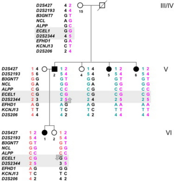
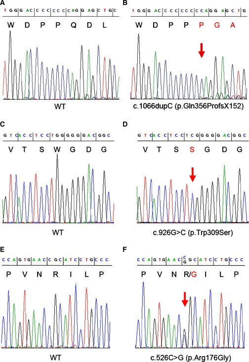
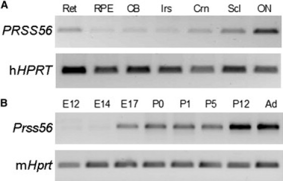
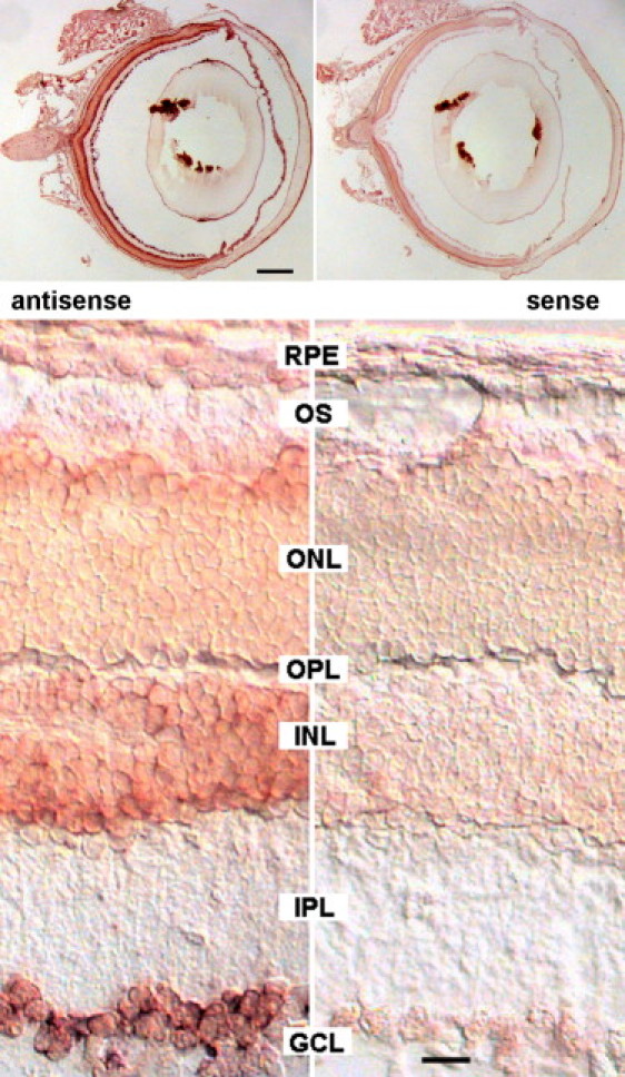
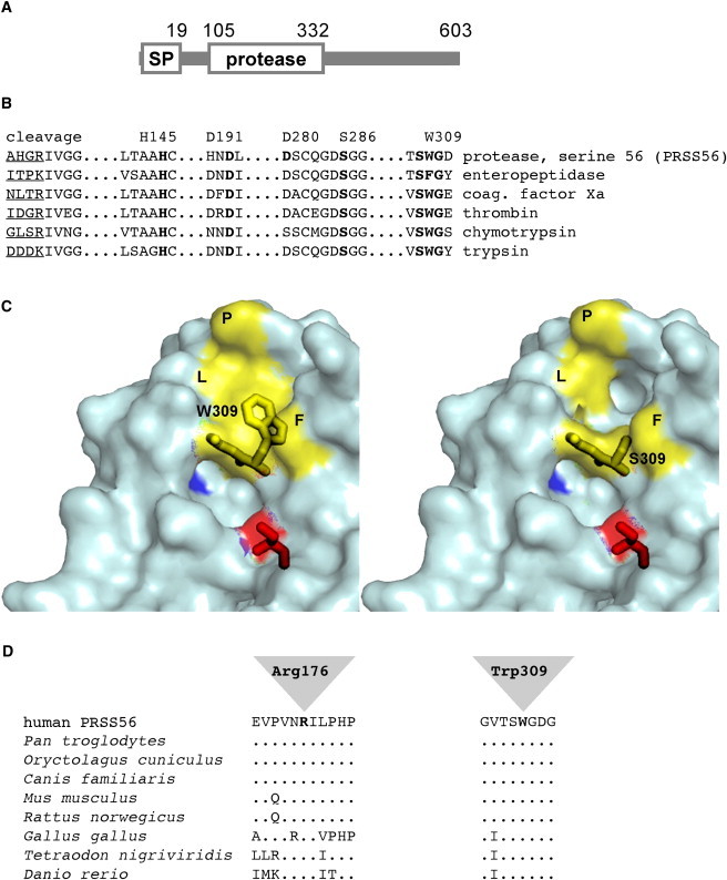
Posterior microphthalmos (MCOP) is a rare isolated developmental anomaly of the eye characterized by extreme hyperopia due to short axial length. The population of the Faroe Islands shows a high prevalence of an autosomal-recessive form (arMCOP) of the disease. Based on published linkage data, we refined the position of the disease locus (MCOP6) in an interval of 250 kb in chromosome 2q37.1 in two large Faroese families. We detected three different mutations in PRSS56. Patients of the Faroese families were either homozygous for c.926G>C (p.Trp309Ser) or compound heterozygous for c.926G>C and c.526C>G (p.Arg176Gly), whereas a homozygous 1 bp duplication (c.1066dupC) was identified in five patients with arMCOP from a consanguineous Tunisian family. In one patient with MCOP from the Faroe Islands and in another one from Turkey, no PRSS56 mutation was detected, suggesting nonallelic heterogeneity of the trait. Using RT-PCR, PRSS56 transcripts were detected in samples derived from the human adult retina, cornea, sclera, and optic nerve. The expression of the mouse ortholog could be first detected in the eye at E17 and was maintained into adulthood. The predicted PRSS56 protein is a 603 amino acid long secreted trypsin-like serine peptidase. The c.1066dupC is likely to result in a functional null allele, whereas the two point mutations predict the replacement of evolutionary conserved and functionally important residues. Molecular modeling of the p.Trp309Ser mutant suggests that both the affinity and reactivity of the enzyme toward in vivo protein substrates are likely to be substantially reduced.


Figures

Figure 1
Figure 1
Mapping the MCOP6 Locus in a Branch of Family HOP000201 by the Analysis of Multiple Informative Meioses Alleles of a total of ten DNA polymorphisms in the region between D2S427 and D2S206 on chromosome 2q37.1 are arranged as probable haplotypes. Individuals V.2 and VI.2 carry recombinant chromosomes, suggesting that the MCOP6 locus maps between ALPP and EFHD1. Generation and individual numbers shown in the figure are the same as in Figure S1.
Figure 2
Figure 2
Electropherograms of DNA Sequences of PCR Amplicons (A–F) The panels show the homozygous c.1066dupC and c.926G>C mutations of PRSS56 in patients NB8 (B) and III.5/HOP000202 (D) and the heterozygous c.526C>G mutation in V.2/HOP000201 (F), as well as the corresponding regions in controls (wild-type, WT; A, C, and E, respectively). DNA and predicted amino acid (one letter code) sequences are shown on the top of the figure.
Figure 3
Figure 3
Expression Analysis (A) RT-PCR products from human eye tissues. The following abbreviations are used: Ret, retina; RPE, retinal pigment epithelium/choroid; CB, ciliary body; Irs, iris; Crn, cornea; Scl, sclera; ON, optic nerve. (B) RT-PCR products from mouse eye and retinas at the ages shown. Total RNA was isolated from human donor (male, 60 yr) eye tissues obtained within 20 hr postmortem. Mouse total RNA was isolated from whole eyes of C57Bl/6 at embryonic day ages E12, E14, and E17 and postnatal day ages P0 and P1, as well as from dissected retinas at ages P5, P12, and adult (Ad). RNA concentrations and integrity were evaluated with Agilent Bioanalysis. First-strand cDNAs were prepared from 1 μg RNA with Superscript II, followed by treatment with RNase H. DNA sequences were PCR amplified with primers for PRSS56, Prss56, and the hypoxanthine phosphoribosyltransferase gene (HPRT; Table S3) by using 35 cycles of 94°C for 1 min, 55°C for 1 min, and 68°C for 2 min. The products were visualized on agarose gels stained with SyberGreen.
Figure 4
Figure 4
In Situ Hybridization of Sections of a 3-Month-Old Balb/c Mouse Eye with Prss56 RNA Probes Sagittal sections were hybridized with digoxygenin-labeled antisense and sense RNA probes transcribed from a 411 bp fragment of the Prss56 cDNA (accession number JF323950; amplified with primer pair 2 from P12 mouse retina RNA; see Table S3 for primers). Probes were detected with alkaline phosphatase-coupled anti-DIG antibodies, followed by staining in Nitro-Blue Tetrazolium/5-Bromo-4-Chloro-3′-Indolyphosphate (NBT/BCIP; Roche) for 4 hr at 37°C. Sections were viewed with a Zeiss Axiophot microscope equipped with differential interference contrast optics and a Kappa camera. Scale bars represent 0.5 mm (top panels) and 20 μm (bottom panels).
Figure 5
Figure 5
Trypsin-Like Serine Protease PRSS56 (A) Schematic structure of PRSS56. Positions of the signal peptide (SP, cleaved between residues 19 and 20), the cleavage site of the proprotein (residues104–105), and the protease domain are indicated. The C-terminal domain of the protein does not exhibit similarity to any known protein. (B) Alignment of functionally relevant sequence elements of members of the serine-protease family. The position of the cleavage site for conversion of the proenzymes into the active enzymes is indicated (trypsinogen to trypsin, prothrombin to thrombin, etc.); the sequence N-terminal to the cleavage site is underlined. Elements of the charge relay system or catalytic triad (Asp191/His145/Ser286 in PRSS56), as well as an aspartate residue localized in the P1 specificity pocket (Asp280 in PRSS56), and the sequence surrounding Trp309 are indicated by bold print. (C) 3D model of wild-type (left) and p.Trp309Ser mutant (right) PRSS56 based on the atomic structure of trypsin. Molecular modeling was performed with the SWISS-MODEL server in automated mode. PDB data sets obtained by the modeling procedure were visualized with the help of the polyview 3D visualization server. Residues Trp309-Gly310 and the catalytic Ser286 (red) are indicated by stick models, and the negatively charged Asp280 at the bottom of the P1 active-site pocket is shown in blue. In the wild-type, Trp309 together with residues Phe188 (F), Leu260 (L), and Pro262 (P) contributes to the formation of a shallow hydrophobic cavity (yellow surface), which is disrupted in the p.Trp309Ser mutant. (D) Sequence alignment of vertebrate homologs of PRSS56. Sequences flanking Arg176 and Trp309 are shown. Identical amino acids are indicated by dots.

References

    1. Fuchs J., Holm K., Vilhelmsen K., Rosenberg T., Scherfig E., Fledelius H.C. Hereditary high hypermetropia in the Faroe Islands. Ophthalmic Genet. 2005;26:9–15. - PubMed
    1. Hmani-Aifa M., Ben Salem S., Benzina Z., Bouassida W., Messaoud R., Turki K., Khairallah M., Rebaï A., Fakhfekh F., Söderkvist P., Ayadi H. A genome-wide linkage scan in Tunisian families identifies a novel locus for non-syndromic posterior microphthalmia to chromosome 2q37.1. Hum. Genet. 2009;126:575–587. - PubMed
    1. Fledelius H.C., Fuchs H.J., Rosenberg T. Oculometric characteristics of extreme hypermetropia in two faroese families. Optom. Vis. Sci. 2004;81:762–768. - PubMed
    1. Serrano J.C., Hodgkins P.R., Taylor D.S., Gole G.A., Kriss A. The nanophthalmic macula. Br. J. Ophthalmol. 1998;82:276–279. - PMC - PubMed
    1. Walsh M.K., Goldberg M.F. Abnormal foveal avascular zone in nanophthalmos. Am. J. Ophthalmol. 2007;143:1067–1068. - PubMed

Publication types

MeSH terms

Associated data