

Comment
. 2011 Mar 11;88(3):392-3; author reply 393-5.
doi: 10.1016/j.ajhg.2010.12.015.
Neurodegenerative disorder related to AIMP1/p43 mutation is not a PMLD
- PMID: 21397067
- PMCID: PMC3059435
- DOI: 10.1016/j.ajhg.2010.12.015
Item in Clipboard
Comment
Neurodegenerative disorder related to AIMP1/p43 mutation is not a PMLD
Am J Hum Genet.
.
No abstract available
Figures
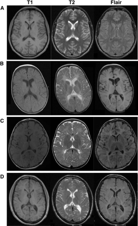
Magnetic Resonance Imaging of White Matter Changes in Hypomyelinating Leukodystrophies (A–D) In a normal brain (A), when the myelination is achieved, the white matter signal is hyperintense relative to gray matter on T1-weighted images and hypointense relative to gray matter on T2-weighted and FLAIR images. In patients affected by hypomyelinating leukodystrophies, T1 white matter signal is hypointense relative to gray matter in severe form 0 related to proteolipid potein 1 (PLP1 [MIM 300401]) mutation (patient B, 2 years old), isointense in moderate form 3 related to PLP1 duplication (patient C, 7 years old), and normally hyperintense in mild form 4 related to gap junction protein gamma-2 (GJC2 [MIM 608803]) mutation (patient D, 20 years old). In patients B and C, the white matter signal on T2-weighted and FLAIR images appears hyperintense relative to gray matter. In patient D, the white matter tends to be isointense relative to gray matter on T2-weighted images, except in posterior internal capsules, and is hyperintense in FLAIR images.
Comment on
-
Pelizaeus-Merzbacher-like disease caused by AIMP1/p43 homozygous mutation.Am J Hum Genet. 2010 Dec 10;87(6):820-8. doi: 10.1016/j.ajhg.2010.10.016. Epub 2010 Nov 18. Am J Hum Genet. 2010. PMID: 21092922 Free PMC article.
References
-
- Mimault C., Giraud G., Courtois V., Cailloux F., Boire J.Y., Dastugue B., Boespflug-Tanguy O., The Clinical European Network on Brain Dysmyelinating Disease Proteolipoprotein gene analysis in 82 patients with sporadic Pelizaeus-Merzbacher Disease: Duplications, the major cause of the disease, originate more frequently in male germ cells, but point mutations do not. Am. J. Hum. Genet. 1999;65:360–369. - PMC - PubMed
-
- Henneke M., Combes P., Diekmann S., Bertini E., Brockmann K., Burlina A.P., Kaiser J., Ohlenbusch A., Plecko B., Rodriguez D. GJA12 mutations are a rare cause of Pelizaeus-Merzbacher-like disease. Neurology. 2008;70:748–754. - PubMed
-
- Boespflug-Tanguy O., Labauge P., Fogli A., Vaurs-Barriere C. Genes involved in leukodystrophies: A glance at glial functions. Curr. Neurol. Neurosci. Rep. 2008;8:217–229. - PubMed
Publication types
MeSH terms
Substances
LinkOut - more resources
Full Text Sources
Medical
Molecular Biology Databases
Miscellaneous
