Review
. 2011 Apr;34(4):177-87.
doi: 10.1016/j.tins.2011.02.001.

Experience-dependent structural plasticity in the cortex

Affiliations
Review

Experience-dependent structural plasticity in the cortex

Min Fu et al. Trends Neurosci. 2011 Apr.

Abstract

Synapses are the fundamental units of neuronal circuits. Synaptic plasticity can occur through changes in synaptic strength, as well as through the addition/removal of synapses. Two-photon microscopy in combination with fluorescence labeling offers a powerful tool to peek into the living brain and follow structural reorganization at individual synapses. Time-lapse imaging depicts a dynamic picture in which experience-dependent plasticity of synaptic structures varies between different cortical regions and layers, as well as between neuronal subtypes. Recent studies have demonstrated that the formation and elimination of synaptic structures happens rapidly in a subpopulation of cortical neurons during various sensorimotor learning experiences, and that stabilized synaptic structures are associated with long lasting memories for the task. Therefore, circuit plasticity, mediated by structural remodeling, provides an underlying mechanism for learning and memory.


Figures

Figure 1
Figure 1
Transcranial two-photon imaging of fluorescently labeled pyramidal cortex neurons in the living mouse brain. (a) This image illustrates a subset of YFP-expressing L5 pyramidal neurons that extend their apical dendrites to L1 in the barrel cortex of a one-month-old YFP-H transgenic mouse. Scale bar: 50 μm. Modified, with permission, from [36]. (b) A CCD camera image of the brain vasculature as observed in the mouse cortex using the thinned skull method of preparation. Scale bar: 1 mm. (c–d) Dendritic spine dynamics of L5 pyramidal neurons in the barrel cortex of a YFP-H line adolescent mouse. (c) A low-magnification 2D projection from a 3D stack of dendritic branches and axons. Scale bar: 10 μm. (d) A higher magnification view of the dendritic segment demarcated in the white box in (c). Two images of the same dendritic segment, taken at the ages of weeks (w) 4 and 6, respectively, reveal spine elimination (green arrowheads), spine formation (red arrowheads) and filopodia (asterisks). Scale bar: 2 μm. (c) and (d) are modified, with permission, from [27]. (e) En passant bouton dynamics of intracortical axons from L2/3 and L4 neurons in the barrel cortex of a GFP-M transgenic mouse. Two images of the same axon segment, taken at P86 and P94, respectively, reveal the addition of an en passant bouton (red arrowhead). Scale bar: 10 μm. (f) Terminaux bouton dynamics of thalmocortical axons in the barrel cortex of a GFP-M transgenic mouse. Two images of the same axon segment, taken on P124 and P128, respectively, reveal the addition of a terminaux bouton (red arrowhead). Scale bar: 5 μm. (e) and (f) are modified, with permission, from [59]. (g) Dendritic tip dynamics of layer 2/3 interneurons in the visual cortex of a GFP-S transgenic mouse. Two images of the same dendrite, taken at the age of weeks 11 and 13, respectively, reveal elongation of a dendritic tip (pointed by 3 red arrowheads). Scale bar: 5 μm. Modified, with permission, from [58].
Figure 2
Figure 2
Spine dynamics and morphological changes associated with learning. (a) Intrinsic dendritic spine turnover correlates with the ability to learn new tasks. In zebra finches (upper panel), the forebrain nucleus HVC is the proximal site where auditory information merges with an explicit song motor representation. Labeled with a lentivirus expressing EGFP, spine turnover of HVC neurons in juvenile songbirds was measured over two hours the night before the first exposure to a song tutor [48]. The scatter plot illustrates that the bird’s capacity of subsequent song imitation correlates with its degree of spine turnover before learning. Each circle represents a single bird. The black line is fitted by linear regression, r=0.63. Modified, with permission, from [48]. (b) Dendritic spine formation during learning correlates with learning performance. YFP-H transgenic mice were trained in a single-pellet reaching task (upper panel). The apical dendrites of L5 neurons in the motor cortex contralateral to the trained limb were imaged the day before and immediately following the first training session. The scatter plot shows that the number of successful reaches during the first training session correlates with the degree of spine formation. Each circle represents one mouse. The black line is fitted by linear regression, r=0.88. Modified, with permission, from [37]. (c) Fractions of stabilized new spines correlate with performance in the re-train session. YFP-H transgenic mice were trained on an accelerating rotarod (upper panel). Dendritic spines of the L5 neurons in motor cortex were imaged at three time points: before training, after 2 days of training, and before re-training on day 7. The scatter plot shows that an animal’s performance in the re-train session on day 7 correlates with stabilized new spines formed in the first 2 days of training. Each circle represents one mouse. The black line is fitted by linear regression, r=0.93. Modified, with permission, from [38]. (d–e) Changes in dendritic spine sizes during visual manipulation. Adolescent GFP-M transgenic mice underwent two 8-day monocular deprivations (MD) interrupted by a 20-day period of binocular vision (BV). Dendritic spines of L5 pyramidal neurons in the binocular region of the visual cortex were imaged every four days. (d) A spine formed during the first four days of MD (red arrowhead) decreases its brightness/size during timepoints within the interval BV period, and then increases its brightness/size during the second period of MD. (e) Average integrated brightness of persistent new spines formed during MD (red) or during the corresponding time period in non-deprived control animals (grey), normalized to the spine brightness value at the end of the first MD or the equivalent time point in control mice. Mean ± s.e.m. Modified, with permission, from [30].
Figure 3
Figure 3
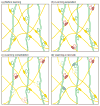
Schematic drawing illustrating spine remodeling and maintenance in the mouse motor cortex when a mouse is trained with different forelimb motor tasks sequentially (based on experimental data from [37]). (a) Three spiny dendrites (green) connect with their neighboring axons (yellow) at 6 different synaptic sites (identified as spines and boutons, respectively). (b) New synaptic connections (red) form rapidly during the acquisition phase of learning the first motor task (i.e., a single-pellet reaching task). (c) During the learning consolidation phase of the same motor task, a majority of new synapses formed during learning acquisition are selectively stabilized, while synapses that existed prior to learning are preferentially eliminated (with dotted outlines). This results in a rewiring of the circuit without overall changes in synaptic density. (d) Learning a different forelimb motor task (i.e., a pasta handling task) later in life remodels a different set of synapses (blue).

References

    1. James W. The Principles of Psychology. Vol. 1 Henry Holt & Co; 1890.
    1. Hubel DH, Wiesel TN. Early exploration of the visual cortex. Neuron. 1998;20:401–412. - PubMed
    1. Hubel DH, Wiesel TN. The period of susceptibility to the physiological effects of unilateral eye closure in kittens. J Physiol. 1970;206:419–436. - PMC - PubMed
    1. Hubel DH, et al. Plasticity of ocular dominance columns in monkey striate cortex. Philos Trans R Soc Lond B Biol Sci. 1977;278:377–409. - PubMed
    1. Wiesel TN, Hubel DH. Single-Cell Responses in Striate Cortex of Kittens Deprived of Vision in One Eye. J Neurophysiol. 1963;26:1003–1017. - PubMed

Publication types