. 2011 May 15;353(2):309-20.
doi: 10.1016/j.ydbio.2011.03.004. Epub 2011 Mar 21.

Conditional expression of the TVA receptor allows clonal analysis of descendents from Cre-expressing progenitor cells

Affiliations

Conditional expression of the TVA receptor allows clonal analysis of descendents from Cre-expressing progenitor cells

Kevin T Beier et al. Dev Biol. .

Abstract

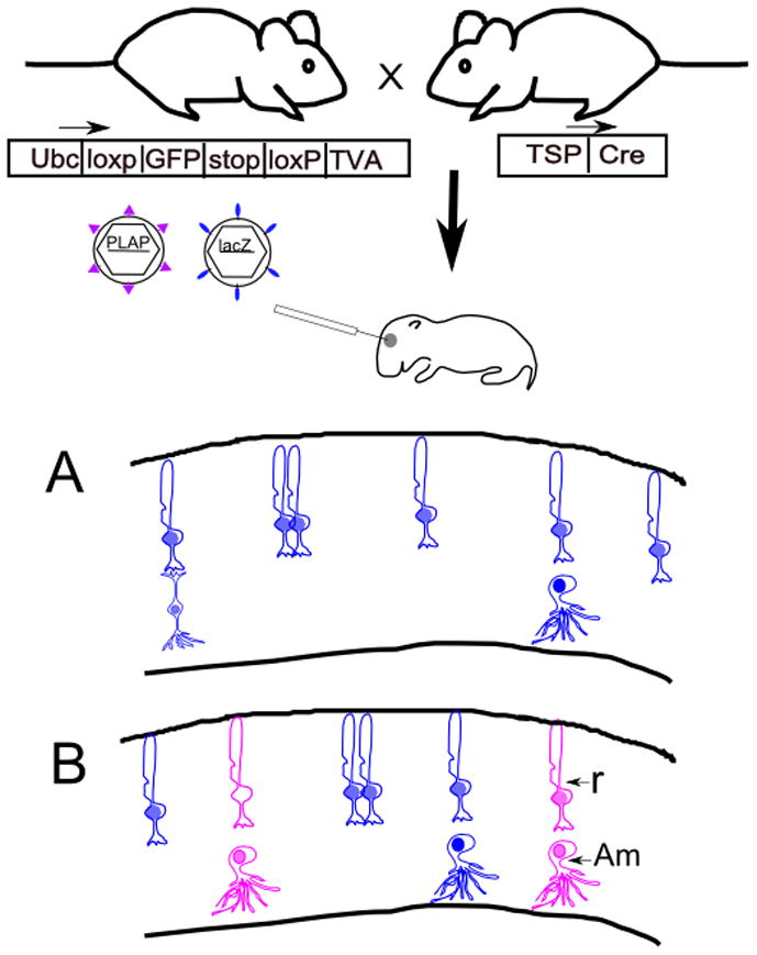
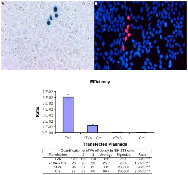
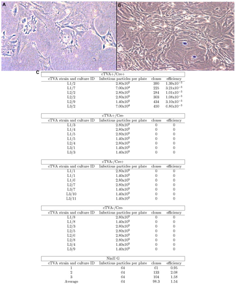
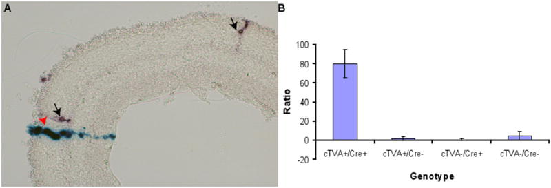
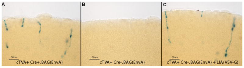
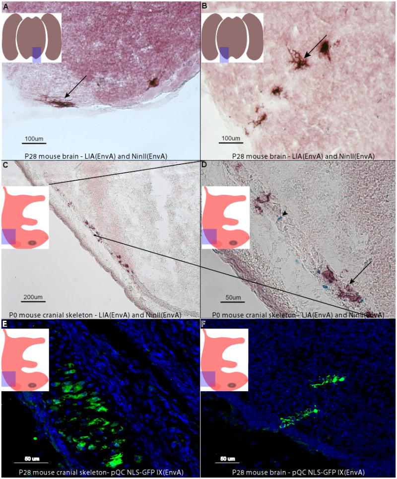
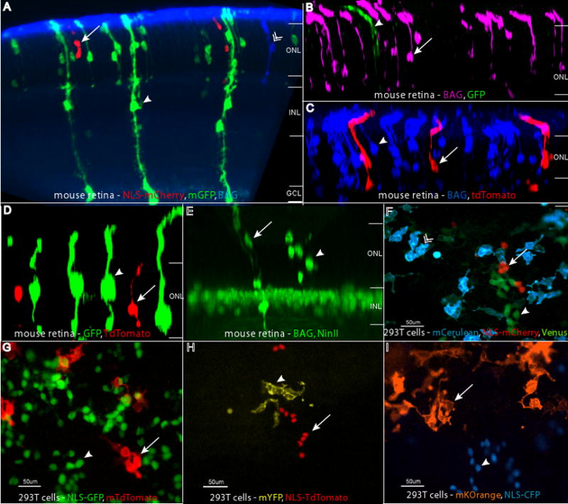
An understanding of the number and types of progeny produced by progenitor cells during development provides a foundation for studies of when and where cell fate determination takes place. Lineal relationships can be revealed by the identification of descendents of cells that express a recombinase, such as Cre or Flp. This method provides data concerning gene expression history, but does not provide clonal resolution among the descendents. An alternative method employs retroviral labeling, which permits the identification of clones, but does not allow for the tracking of gene expression history. Here we report a combination of these methods to circumvent each method's limitations. By employing the specificity of Cre expression, and by selecting only a subset of cells with a Cre history for retroviral infection, clones with a gene expression history can be labeled. The method utilizes a conditional allele of the avian tumor virus receptor A (TVA), which allows infection of mouse cells following Cre activity, with mammalian retroviral vectors pseudotyped with the ASLV-A envelope glycoprotein (EnvA). We quantified the efficiency and specificity of this system in vivo and in vitro. We also generated a series of retroviral vectors encoding a variety of histochemical and fluorescent reporter genes that enable the tracking of mixtures of clones, thus enabling better resolution of clonal boundaries. This method and new vectors can be used to further our understanding of the gene expression patterns of progenitor cells that make particular daughter cells, as well as provide a platform for manipulating identified subsets of developing cells.


Figures

Figure 1
Figure 1
Experimental strategy for use of the cTVA allele for lineage analysis. A mouse with conditional expression of TVA can be crossed to a Cre line of choice, with expression of Cre directed by a tissue-specific promoter (TSP). TVA, a receptor for viruses with the EnvA glycoprotein, will be expressed in cells with a history of Cre expression. Viruses pseudotyped with the EnvA glycoprotein are able to infect TVA-expressing cells, and are not able to infect non-TVA expressing mouse cells. Viruses pseudotyped with the VSV-G glycoprotein can infect any mitotic cell. Two viruses, one pseudotyped with EnvA, encoding PLAP, and one pseudotyped with VSV-G, encoding lacZ, can be coinjected into a developing tissue, e.g. the eye. The drawings in A and B depict retinal sections following histochemical processing for PLAP (purple) and β-galactosidase (blue). (A). If a control, TVA-negative retina is injected with a mixture of the two viruses, only blue clones will result. (B) If a TVA expressing retina is injected with this mixture of viruses, both types of viruses will infect mitotic cells, giving rise to blue and purple clones. In this example, the cells with a Cre history generate clones of rod photoreceptors (R) and amacrine interneurons (Am).
Figure 2
Figure 2
Infection of murine NIH 3T3 cells with EnvA pseudotyped retroviruses. Mouse NIH 3T3 cells were transfected with either pCAG-TVA, pUbC-cTVA + pCAG-GFP:Cre, pUbC-cTVA, or CAG-GFP:Cre. The next day, they were infected with MMLV retroviruses pseudotyped with the EnvA glycoprotein. (A) An image of a single NinII-infected clone detected by Xgal histochemistry. (B) An image of a single fluorescent NLS-tdTomato-infected clone (red) with DAPI-labeled nuclei (blue). The infection ratios, which are the number of clones observed on the transfected cells vs. the number of infectious particles observed on 293T TVA 800 cells (which constitutively express TVA, (Narayan et al., 2003)) are plotted with corresponding values shown in the table below. The number of infections in the cTVA alone or CAG-GFP:Cre alone conditions were not significantly different from each other (t-test).
Figure 3
Figure 3
Infection of cTVA MEFs with EnvA pseudotyped retroviruses. MEFS from three different cTVA mouse lines (L1, L2, and L3) were infected with NinII(EnvA). (A) An image of a dish from a TVA- Cre- culture. (B) An image from a dish of a TVA+/Cre+ culture. Both cultures were infected with the same number of NinII infectious particles. (C) Quantitative data from the infection of MEFs by NinII. Column 1 lists the culture ID number, which refers to an individual MEF culture, derived from a unique embryo. Column 2 indicates the number of infectious viral particles applied to the dish. Column 3 indicates the number of clones observed. Column 4 is the ratio of clones observed (column 3) divided by the infectious virions applied (column 2). The number of infectious virions was measured by infection of 293 TVA800 cells, which constitutively express TVA.
Figure 4
Figure 4
Efficiency of infection of virus pseudotyped with EnvA vs. VSV-G. Retroviruses (LIA(EnvA) and BAG (VSV-G)) were mixed and injected into the subretinal space of P0 mice in vivo. Retinas were analyzed for clones following the completion of development at P21, either by scoring infections on retinal whole mount preparations, or by scoring clones on cryosections. (A) A cryosection of a retina exhibiting clones from LIA(EnvA) (arrows) and BAG (VSV-G) (arrowhead) are shown. (B) The ratio of VSV-G to EnvA events was calculated for the four resulting genotypes of the cTVA+/− x hβactin-Cre +/− cross, as shown. The values for these calculations are listed in Table 1.
Figure 5
Figure 5
Test for effect of a virus with one glycoprotein on the infectivity of a virus with a different glycoprotein. EnvA retroviruses were used to infect retinal explants with and without co-infection with VSV-G retroviruses. Retinal explants were infected with BAG(EnvA) at P0. The retina in (A) was TVA+/Cre+ and was infected with BAG(EnvA) retrovirus only, while that in (B) was TVA+/Cre− and was infected with the same amount of infectious BAG(EnvA) virus. The retina in (C) was also TVA+/Cre−, but was infected with a mixture of BAG(EnvA) and LIA(VSV-G). All retinae were stained for β-galactosidase only.
Figure 6
Figure 6
Infection of mouse brains with LIA and NinII pseudotyped with EnvA. P2 mouse pups of the cross cTVA+/− x hβactin-Cre +/− were injected in the subventricular zone of the midbrain and sacrificed at P28. Coronal sections were made and stained histochemically for PLAP and β-galactosidase activity. The area of the sections is indicated in the highlighted area of the embedded cartoon. Both viruses labeled oligodendrocytes (A, arrow) and astrocytes (B, arrow), glial types known to be produced postnatally. Viral labeling was only seen in animals with TVA and Cre; all other genotypes of this cross were negative. Wnt1-Cre +/− mice were crossed to cTVA +/− mice, and the embryos were injected in utero into the third ventricle with LIA(EnvA) and NinII(EnvA), or QC NLS-GFP IX(EnvA) (C–F). The embedded cartoons show the area of the sections that are shown. Cells in the cranial skeleton that are descended from Wnt1 expressing progenitor cells, infected by LIA(EnvA) (arrow in D), NinII(EnvA) (arrowhead in D), or QC NLS-GFP IX(EnvA) (E) in cTVA+/Cre+ mice, are shown. Hindbrain neurons also were labeled, as demonstrated by a labelled Purkinje cell (F), generated by infection with QC NLS-GFP IX(EnvA). Injections into TVA− or Cre− animals were negative.
Figure 7
Figure 7
Retroviruses with distinct histochemical and fluorescent reporters. Combinations of BAG, LIA, and QC viruses were injected in vivo into the subretinal space of P0 retinas, and the retinas were examined at P21. The reporter combinations shown are (A) GFP (anti-GFP, in green, arrowhead), mCherry (anti-RFP, in red, arrow), and BAG (anti-β-galactosidase, in blue, chevron), (B), GFP (anti-GFP, in green, arrowhead) and BAG (anti-β-galactosidase, in magenta, arrow) (C), tdTomato (anti-RFP, in red, arrow) and BAG (anti-β-galactosidase, in blue, arrowhead), (D) GFP (anti-GFP, in green, arrowhead) and tdTomato (anti-RFP, in red, arrow), (E) NinII (anti-β-gal, in green, arrowhead) and BAG (anti-β-gal, in green, arrow) with Chx10-NLS-GFP, a gene in the transgenic background, marking bipolar cell nuclei. The retinal layers are labeled in panels A–E as follows: ONL = outer nuclear layer, INL = inner nuclear layer, GC = ganglion cell layer. Panels F–I show images of infected 293T cells. These combinations are: (F) membrane Cerulean (blue, chevron), Venus (green, arrowhead), and NLS-mCherry (red, arrow), (G) NLS-GFP (green, arrowhead) and membrane tdTomato (red, arrow), (H) NLS-tdTomato (red, arrow) and membrane YFP (yellow, arrowhead), and (I) membrane Kusabira Orange (orange, arrow) and NLS-CFP (blue, arrowhead).

References

    1. Altman J. Proliferation and migration of undifferentiated precursor cells in the rat during postnatal gliogenesis. Exp Neurol. 1966;16:263–278. - PubMed
    1. Altshuler D, Lo Turco JJ, Rush J, Cepko C. Taurine promotes the differentiation of a vertebrate retinal cell type in vitro. Development. 1993;119:1317–1328. - PubMed
    1. Bao ZZ, Cepko CL. The expression and function of Notch pathway genes in the developing rat eye. J Neurosci. 1997;17:1425–1434. - PMC - PubMed
    1. Bates P, Young JA, Varmus HE. A receptor for subgroup A Rous sarcoma virus is related to the low density lipoprotein receptor. Cell. 1993;74:1043–1051. - PubMed
    1. Battini JL, Rodrigues P, Müller R, Danos O, Heard JM. Receptor-binding properties of a purified fragment of the 4070A amphotropic murine leukemia virus envelope glycoprotein. J Virol. 1996;70:4387–4393. - PMC - PubMed

Publication types

MeSH terms

LinkOut - more resources